Ultrasound
Diagnose causes of pain, swelling, and infection
General Ultrasound
Ultrasound imaging uses sound waves to produce pictures of the inside of the body. It is used to help diagnose the causes of pain, swelling and infection in the body’s internal organs and to examine a baby in pregnant women. It’s also used to help guide biopsies, diagnose heart conditions, and assess damage after a heart attack. Ultrasound is safe, noninvasive, and does not use ionizing radiation.
This procedure requires little to no special preparation. Your doctor will instruct you on how to prepare, including whether you should refrain from eating or drinking beforehand. Leave jewelry at home and wear loose, comfortable clothing. You may be asked to wear a gown.
What is General Ultrasound Imaging?
Ultrasound is safe and painless. It produces pictures of the inside of the body using sound waves. Ultrasound imaging is also called ultrasound scanning or sonography. It uses a small probe called a transducer and gel placed directly on the skin. High-frequency sound waves travel from the probe through the gel into the body. The probe collects the sounds that bounce back. A computer uses those sound waves to create an image. Ultrasound exams do not use radiation (as used in x-rays). Because images are captured in real-time, they can show the structure and movement of the body’s internal organs. They can also show blood flowing through blood vessels.
Ultrasound imaging is a noninvasive medical test that helps physicians diagnose and treat medical conditions.
Conventional ultrasound displays the images in thin, flat sections of the body. Advancements in ultrasound technology include three-dimensional (3-D) ultrasound that formats the sound wave data into 3-D images.
A Doppler ultrasound study may be part of an ultrasound examination.
Doppler ultrasound is a special ultrasound technique that evaluates movement of materials in the body. It allows the doctor to see and evaluate blood flow through arteries and veins in the body.
There are three types of Doppler ultrasound:
- Color Doppler uses a computer to convert Doppler measurements into an array of colors to show the speed and direction of blood flow through a blood vessel.
- Power Doppler is a newer technique that is more sensitive than color Doppler and capable of providing greater detail of blood flow, especially when blood flow is little or minimal. Power Doppler, however, does not help the radiologist determine the direction of blood flow, which may be important in some situations.
- Spectral Doppler displays blood flow measurements graphically, in terms of the distance traveled per unit of time, rather than as a color picture. It can also convert blood flow information into a distinctive sound that can be heard with every heartbeat.
What are some common uses of the procedure?
Ultrasound examinations can help to diagnose a variety of conditions and to assess organ damage following illness.
Ultrasound is used to help physicians evaluate symptoms such as:
- pain
- swelling
- infection
- mass
- abnormal labs
- hypertension
Ultrasound Exams We Do:
- Breast
- Breast Biopsy
- Abdominal (liver, gallbladder, kidney, aorta, spleen, pancreas)
- Renal (Kidney & Bladder)
- Thyroid/Parathyroid
- Pelvic
- Soft Tissue (Palpable masses any place on body) Lymph Nodes
- Chest
- Fetal Age (OB’s)
- Carotid
- Renal Artery Duplex
- Scrotal
- Hernia Sono
- Venous Duplex
Doppler ultrasound images can help the physician to see and evaluate:
- blockages to blood flow (such as clots)
- narrowing of vessels
- tumors and congenital vascular malformations
- reduced or absent blood flow to various organs, such as the testes or ovary
- increased blood flow, which may be a sign of infection
With knowledge about the speed and volume of blood flow gained from a Doppler ultrasound image, the physician can often determine whether a patient is a good candidate for a procedure like angioplasty.
How should I prepare?
Wear comfortable, loose-fitting clothing. You may need to remove all clothing and jewelry in the area to be examined.
You may be asked to wear a gown during the procedure.
Preparation for the procedure will depend on the type of examination you will have. For some scans your doctor may instruct you not to eat or drink for as many as 12 hours before your appointment. For others you may be asked to drink up to six glasses of water two hours prior to your exam and avoid urinating so that your bladder is full when the scan begins.
What does the equipment look like?
Ultrasound scanners consist of a computer console, video display screen and an attached transducer. The transducer is a small hand-held device that resembles a microphone. Some exams may use different transducers (with different capabilities) during a single exam. The transducer sends out inaudible, high-frequency sound waves into the body and then listens for the returning echoes. The principles are similar to sonar used by boats and submarines.
The technologist applies a small amount of gel to the area under examination and places the transducer there. The gel allows sound waves to travel back and forth between the transducer and the area under examination. The ultrasound image is immediately visible on a video display screen that looks like a computer monitor. The computer creates the image based on the loudness (amplitude), pitch (frequency) and time it takes for the ultrasound signal to return to the transducer. It also takes into account what type of body structure and/or tissue the sound is traveling through.
How does the procedure work?
Ultrasound imaging is based on the same principles involved in the sonar used by bats, ships and fishermen. When a sound wave strikes an object, it bounces back, or echoes. By measuring these echo waves, it is possible to determine how far away the object is as well as the object’s size, shape and consistency. This includes whether the object is solid or filled with fluid.
In medicine, ultrasound is used to detect changes in the appearance of organs, tissues, and vessels and to detect abnormal masses, such as tumors.
In an ultrasound exam, a transducer both sends the sound waves and records the echoing waves. When the transducer is pressed against the skin, it sends small pulses of inaudible, high-frequency sound waves into the body. As the sound waves bounce off internal organs, fluids and tissues, the sensitive receiver in the transducer records tiny changes in the sound’s pitch and direction. These signature waves are instantly measured and displayed by a computer, which in turn creates a real-time picture on the monitor. One or more frames of the moving pictures are typically captured as still images. Short video loops of the images may also be saved.
Doppler ultrasound, a special ultrasound technique, measures the direction and speed of blood cells as they move through vessels. The movement of blood cells causes a change in pitch of the reflected sound waves (called the Doppler effect). A computer collects and processes the sounds and creates graphs or color pictures that represent the flow of blood through the blood vessels.
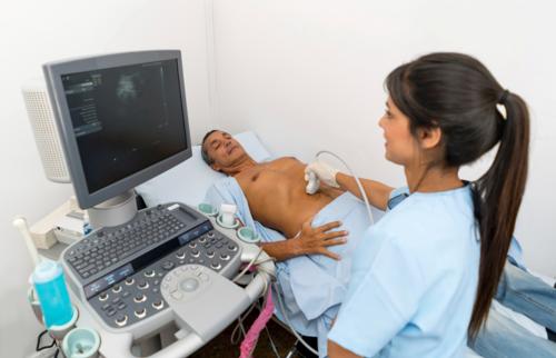
How is the procedure performed?
For most ultrasound exams, you will lie face-up on an exam table that can be tilted or moved.
Patients may be turned to either side to improve the quality of the images.
After you are positioned on the examination table, the radiologist (a physician specifically trained to supervise and interpret radiology examinations) or sonographer will apply a warm water-based gel to the area of the body being studied. The gel will help the transducer make secure contact with the body and eliminate air pockets between the transducer and the skin that can block the sound waves from passing into your body. The transducer is placed on the body and moved back and forth over the area of interest until the desired images are captured.
There is usually no discomfort from pressure as the transducer is pressed against the area being examined. However, if scanning is performed over an area of tenderness, you may feel pressure or minor pain from the transducer.
Doppler sonography is performed using the same transducer.
Rarely, young children may need to be sedated in order to hold still for the procedure. Parents should ask about this beforehand and be made aware of food and drink restrictions that may be needed prior to sedation.
Once the imaging is complete, the clear ultrasound gel will be wiped off your skin. Any portions that are not wiped off will dry quickly. The ultrasound gel does not usually stain or discolor clothing.
In some ultrasound studies, the transducer is attached to a probe and inserted into a natural opening in the body.
What will I experience during and after the procedure?
Most ultrasound exams are painless, fast and easily tolerated.
Ultrasound exams in which the transducer is inserted into an opening of the body may produce minimal discomfort.
If a Doppler ultrasound study is performed, you may actually hear pulse-like sounds that change in pitch as the blood flow is monitored and measured.
Most ultrasound examinations are completed within 30 minutes, although more extensive exams may take up to an hour.
When the examination is complete, you may be asked to dress and wait while the ultrasound images are reviewed.
After an ultrasound examination, you should be able to resume your normal activities immediately.
Who interprets the results and how do I get them?
A radiologist, a doctor trained to supervise and interpret radiology exams, will analyze the images. The radiologist will send a signed report to the doctor who requested the exam. Your doctor will then share the results with you. In some cases, the radiologist may discuss results with you after the exam.
Follow-up exams may be needed. If so, your doctor will explain why. Sometimes a follow-up exam is done because a potential abnormality needs further evaluation with additional views or a special imaging technique. A follow-up exam may also be done to see if there has been any change in an abnormality over time. Follow-up exams are sometimes the best way to see if treatment is working or if an abnormality is stable or has changed.
What are the benefits vs. risks?
Benefits
- Most ultrasound scanning is noninvasive (no needles or injections).
- Occasionally, an ultrasound exam may be temporarily uncomfortable, but it should not be painful.
- Ultrasound is widely available, easy-to-use and less expensive than most other imaging methods.
- Ultrasound imaging is extremely safe and does not use radiation.
- Ultrasound scanning gives a clear picture of soft tissues that do not show up well on x-ray images.
- Ultrasound is the preferred imaging modality for the diagnosis and monitoring of pregnant women and their unborn babies.
- Ultrasound provides real-time imaging, making it a good tool for guiding minimally invasive procedures such as needle biopsies and fluid aspiration.
Risks
- Standard diagnostic ultrasound has no known harmful effects on humans.
What are the limitations of General Ultrasound Imaging?
Ultrasound waves are disrupted by air or gas. Therefore, ultrasound is not an ideal imaging technique for the air-filled bowel or organs obscured by the bowel. Ultrasound is not as useful for imaging air-filled lungs, but it may be used to detect fluid around or within the lungs. Similarly, ultrasound cannot penetrate bone, but may be used for imaging bone fractures or for infection surrounding a bone.
Large patients are more difficult to image by ultrasound because greater amounts of tissue attenuate (weaken) the sound waves as they pass deeper into the body and need to be returned to the transducer for analysis.
Ultrasound has difficulty penetrating bone and, therefore, can only see the outer surface of bony structures and not what lies within (except in infants who have more cartilage in their skeletons than older children or adults). For visualizing internal structure of bones or certain joints, other imaging modalities such as MRI are typically used.