Magnetic Resonance Imaging (MRI) is a medical imaging technique that uses strong magnetic fields and radio waves to generate detailed images of the body’s internal structures. MRI technology has revolutionized medical imaging, allowing doctors to diagnose and treat a wide range of medical conditions with great accuracy and precision. Since its development in the 1970s, MRI technology has continued to evolve, with advancements in imaging speed, resolution, patient comfort, and safety. In this article, we have provided an overview of the different types of MRI machines, including their advantages and disadvantages, as well as a detailed review of the safety of MRI technology and best practices for ensuring patient safety. We have also discussed potential developments in MRI technology that may emerge in the future, including faster imaging times, higher resolution imaging, improved patient comfort, more specialized imaging, portable MRI machines, improved safety, and the use of artificial intelligence. As MRI technology continues to evolve, it is likely to become an even more important tool for diagnosing and treating a wide range of medical conditions.
Closed MRI
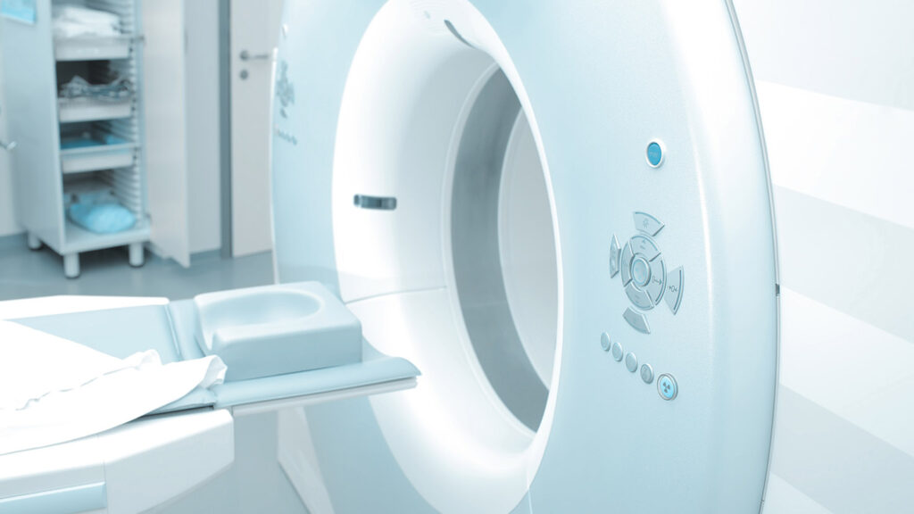
Closed MRI machines, also known as traditional MRI machines, are the most used type of MRI machine. They consist of a narrow tunnel-like structure that the patient lies inside while the machine generates a strong magnetic field and radio waves to create images of the body’s internal structures.
Advantages:
High quality images with excellent spatial resolution: Closed MRI machines produce high-quality images that can help doctors diagnose a wide range of medical conditions.
Wide availability: Closed MRI machines are available at most imaging centers and hospitals.
Can be used to image almost any part of the body: Closed MRI machines are versatile and can be used to image a wide range of anatomical structures.
Disadvantages:
Patients may experience claustrophobia or anxiety due to the narrow tunnel-like structure: Some patients may find the enclosed space uncomfortable or frightening, which can make it difficult to complete the imaging process.
Some patients may be too large to fit inside the machine: Patients who are overweight or who have a large body frame may not be able to fit inside the narrow tunnel of a closed MRI machine.
The machine can be quite loud, which can be uncomfortable or frightening for some patients: The machine produces loud knocking and thumping noises during imaging, which can be unsettling for some patients.
Open MRI
Open MRI machines are designed with a larger opening and can accommodate patients who are claustrophobic or have difficulty lying in a closed space. The open design of the machine may sacrifice some image quality, but it offers greater patient comfort.
Advantages:
Patients with claustrophobia or anxiety can be more comfortable due to the open design of the machine: The open structure of the machine can help patients feel more relaxed during the imaging process.
The machine is less noisy than a closed MRI machine: Open MRI machines produce less noise than traditional MRI machines, which can make the imaging process more comfortable for patients.
The open design can be helpful for patients who are overweight or have mobility issues: The larger opening of an open MRI machine can accommodate patients who may not fit inside a closed MRI machine.
Disadvantages:
Image quality is generally not as good as a closed MRI due to the weaker magnetic field: Open MRI machines use a weaker magnetic field than traditional MRI machines, which can result in lower quality images.
The machine may not be available at all imaging centers: Open MRI machines are not as widely available as traditional MRI machines, which may limit patient access in certain geographic regions.
The open design may not be suitable for all types of imaging, as certain body parts may need to be immobilized to get clear images: The open structure of the machine may not be appropriate for imaging certain anatomical structures.
High-field MRI
High-field MRI machines generate a stronger magnetic field than traditional MRI machines, which results in higher quality images. These machines are used for diagnosing complex medical conditions and researching various diseases.
Advantages:
High-quality images with excellent spatial resolution and contrast: High-field MRI machines can produce detailed images that can help doctors diagnose complex medical conditions.
Useful for diagnosing complex medical conditions and for research purposes: High-field MRI machines are particularly useful for studying the brain and for diagnosing diseases that may be difficult to detect with other imaging methods.
Can image a wide range of anatomical structures and functions: High-field MRI machines can be used to image a wide range of anatomical structures and functions, including the brain, spine, and joints.
Disadvantages:
The stronger magnetic field can be more uncomfortable for some patients: The stronger magnetic field used in high-field MRI machines may cause discomfort or anxiety for some patients.
Patients with certain types of medical devices, such as pacemakers or cochlear implants, may not be able to undergo imaging in a high-field MRI machine: The stronger magnetic field used in high-field MRI machines can interfere with some types of medical devices, making it unsafe for certain patients to undergo imaging.
The machine is larger and more expensive than lower field MRI machines: High-field MRI machines are more expensive to purchase and maintain than lower field MRI machines. They also require more space in the imaging center or hospital.
Low-field MRI
Low-field MRI machines generate a weaker magnetic field than traditional MRI machines, which results in lower quality images. These machines are typically used for diagnosing musculoskeletal and joint problems.
Advantages:
Can be useful for diagnosing musculoskeletal and joint problems: Low-field MRI machines can produce detailed images of the bones and joints, which can help doctors diagnose injuries or diseases affecting these structures.
More affordable and easier to maintain than high-field MRI machines: Low-field MRI machines are less expensive to purchase and maintain than high-field MRI machines.
May be less uncomfortable for some patients due to the weaker magnetic field: The weaker magnetic field used in low-field MRI machines may be more comfortable for some patients, particularly those who experience anxiety or discomfort in an enclosed space.
Disadvantages:
Lower image quality than high-field MRI machines: Low-field MRI machines cannot produce images of the same quality as high-field MRI machines, which may limit their usefulness in some diagnostic applications.
May not be suitable for all types of imaging due to the weaker magnetic field: Low-field MRI machines may not be appropriate for imaging certain anatomical structures, particularly those that require high spatial resolution.
Not as widely available as closed or high-field MRI machines: Low-field MRI machines are less commonly used than traditional or high-field MRI machines, which may limit patient access to this technology.
Standing MRI
Standing MRI machines allow patients to stand or sit during the imaging process. This type of machine is particularly useful for diagnosing joint problems that may be affected by weight-bearing activity.
Advantages:
Allows for imaging of joints under weight-bearing conditions, which can be helpful in diagnosing certain types of joint problems: Standing MRI machines can produce images of the joints while the patient is standing or sitting, which can help diagnose conditions that may not be apparent in traditional MRI images.
Can be more comfortable for patients who cannot lie down for extended periods of time: Patients who cannot lie down comfortably for an extended period of time may find a standing MRI machine more comfortable and easier to tolerate.
Allows for more natural movement during imaging: Patients can move more naturally during imaging in a standing MRI machine, which may help produce more accurate images of joint motion.
Disadvantages:
Image quality may not be as good as a traditional MRI due to the need for patients to move or stand during imaging: Movement during imaging may result in lower quality images, particularly for structures that require high spatial resolution.
The machine may not be widely available at all imaging centers: Standing MRI machines are less commonly used than traditional or high-field MRI machines, which may limit patient access to this technology.
Patients may not be able to stand for the duration of the imaging process: Patients with mobility issues or certain medical conditions may not be able to stand or sit for the duration of the imaging process.
Portable MRI
Portable MRI machines are compact, mobile units that can be transported to different locations. These machines are particularly useful for imaging patients who are unable to travel to a hospital or medical facility.
Advantages:
Can be transported to remote locations, such as rural areas or disaster zones, where traditional MRI machines may not be available: Portable MRI machines can be transported to areas where traditional MRI machines are not available, which can help provide medical imaging to underserved populations or in emergency situations.
Useful for imaging patients who cannot travel to a hospital or medical facility: Portable MRI machines can be used to image patients who are unable to travel to a hospital or medical facility, which can help improve access to medical care for some patients.
Can be less intimidating than a traditional MRI machine: Portable MRI machines can be less intimidating than traditional MRI machines, particularly for patients who are anxious or fearful of medical procedures.
Disadvantages:
Lower image quality than traditional MRI machines: Portable MRI machines may not be able to produce images of the same quality as traditional MRI machines, particularly for structures that require high spatial resolution.
The machine may not be suitable for all types of imaging: Portable MRI machines may not be appropriate for imaging certain anatomical structures or functions, particularly those that require high magnetic field strength or long imaging times.
May be more expensive to use due to the need to transport the machine and set it up at each location: The cost of transporting and setting up a portable MRI machine may be higher than using a traditional MRI machine at a fixed location.
Functional MRI
Functional MRI machines are used to image the brain and monitor changes in blood flow and oxygen levels. These machines can help researchers study brain function and diagnose certain conditions, such as tumors and brain injuries.
Advantages:
Can be used to study brain function and diagnose certain conditions, such as tumors and brain injuries: Functional MRI machines can help doctors diagnose and monitor certain conditions affecting the brain and its function.
Non-invasive and does not require the use of contrast agents: Functional MRI machines are non-invasive and do not require the use of contrast agents, which can be safer for some patients.
Can image both the structure and function of the brain: Functional MRI machines can produce images of both the structure and function of the brain, which can provide a more complete picture of brain health.
Disadvantages:
Lower spatial resolution than traditional MRI machines: Functional MRI machines may not be able to produce images of the same quality as traditional MRI machines, particularly for structures that require high spatial resolution.
Can be more expensive than other types of MRI machines: Functional MRI machines may be more expensive to use than other types of MRI machines, particularly for research purposes.
May not be suitable for imaging other parts of the body besides the brain: Functional MRI machines are designed specifically for imaging the brain and may not be appropriate for imaging other anatomical structures or functions create a detailed view of the future of MRI technology.
Magnetic Resonance Imaging (MRI) technology has come a long way since it was first developed in the 1970s. Today, MRI machines are used to diagnose a wide range of medical conditions, from brain and spinal cord injuries to musculoskeletal disorders and cancer. As technology continues to evolve, we can expect to see significant advancements in MRI technology in the future.
Potential MRI Developments We May See in the Years Ahead
Faster Imaging Times: One area of MRI technology that is likely to see significant improvements in the future is imaging time. Currently, MRI scans can take anywhere from a few minutes to an hour or more, depending on the type of imaging being performed. In the future, we may see advancements in MRI technology that allow for faster imaging times, which could help reduce patient anxiety and improve patient comfort during imaging.
Higher Resolution Imaging: Another area of MRI technology that is likely to see significant advancements in the future is imaging resolution. While current MRI machines produce high-quality images, there is always room for improvement. In the future, we may see advancements in MRI technology that allow for higher resolution imaging, which could help doctors diagnose medical conditions with even greater accuracy.
Improved Patient Comfort: One of the main challenges with MRI imaging is patient comfort. Many patients find the experience uncomfortable, particularly if they are required to lie still in a narrow, enclosed space for an extended period. In the future, we may see advancements in MRI technology that improve patient comfort, such as open MRI machines, which have a larger opening and are less confining than traditional MRI machines.
More Specialized Imaging: Currently, MRI machines are used to diagnose a wide range of medical conditions, but the technology is not always optimized for each specific condition. In the future, we may see advancements in MRI technology that allow for more specialized imaging, such as imaging for specific types of cancer or neurological disorders.
Portable MRI Machines: Another potential development in MRI technology is the development of portable MRI machines. Portable MRI machines could be used to provide medical imaging in remote locations, disaster zones, or in areas where traditional imaging facilities are not available. This could be particularly useful in developing countries, where access to medical care is often limited.
Improved Safety: While MRI technology is generally considered safe, there are some risks associated with the use of strong magnetic fields and radio waves. In the future, we may see advancements in MRI technology that improve safety, such as the development of MRI machines that use weaker magnetic fields, or the use of new imaging agents that are less likely to cause adverse reactions.
Artificial Intelligence (AI): One of the most promising areas of MRI technology is the use of artificial intelligence (AI) to analyze MRI images. AI could be used to help doctors diagnose medical conditions more accurately and quickly, or to identify subtle changes in tissue structure or function that might be missed by human observers.
Overall, the future of MRI technology looks promising, with potential advancements in speed, resolution, patient comfort, safety, and specialization. As these advancements continue to emerge, we can expect to see MRI technology become an even more important tool for diagnosing and treating a wide range of medical conditions.
MRI technology is generally considered to be a safe and non-invasive imaging method. However, as with any medical procedure, there are some risks associated with the use of MRI technology. In this section, we will provide a detailed review of the safety of MRI technology, including the potential risks and best practices for ensuring patient safety.
Potential Risks of MRI Technology
Magnetic fields: One of the main risks associated with MRI technology is exposure to strong magnetic fields. These fields can interfere with certain types of medical devices, such as pacemakers or cochlear implants, which can be dangerous for patients who have these devices.
Radio waves: MRI technology uses radio waves to generate images of the body’s internal structures. While the levels of radio waves used in MRI imaging are generally considered safe, there is some concern that long-term exposure to radio waves could have adverse health effects.
Contrast agents: Some MRI imaging procedures use contrast agents, which are substances that help improve the visibility of certain tissues or structures in the body. While these agents are generally considered safe, they can cause allergic reactions or other adverse reactions in some patients.
Best Practices for Ensuring Patient Safety
Screening for medical devices: Patients who are scheduled for an MRI procedure should be screened for medical devices, such as pacemakers or cochlear implants, that may be affected by the strong magnetic fields used in MRI imaging. If a patient has a medical device that could be affected by MRI technology, alternative imaging methods may be used to ensure patient safety.
Educating patients: Patients should be educated about the potential risks and benefits of MRI imaging, as well as any specific safety measures that may be necessary for their particular imaging procedure.
Monitoring patients during imaging: Patients should be monitored closely during MRI imaging to ensure that they are not experiencing any adverse effects, such as dizziness or discomfort.
Using appropriate imaging protocols: MRI imaging protocols should be tailored to each individual patient, based on their medical history, age, and other factors that may affect their safety and comfort during the imaging procedure.
Using appropriate contrast agents: MRI contrast agents should be selected based on their safety and efficacy, and patients should be screened for any allergies or other contraindications before the contrast agent is administered.
Maintaining and calibrating MRI machines: MRI machines should be regularly maintained and calibrated to ensure that they are functioning properly and that patients are not exposed to excessive magnetic fields or radio waves.
In addition to these best practices, there are also specific safety guidelines and regulations that govern the use of MRI technology. These guidelines may vary by country or region, but generally include requirements for equipment maintenance, patient screening, and personnel training.
Overall, MRI technology is generally considered to be a safe and non-invasive imaging method. However, it is important to be aware of the potential risks and best practices for ensuring patient safety during MRI imaging procedures. By following appropriate safety guidelines and protocols, healthcare providers can help ensure that patients receive the benefits of MRI imaging without any adverse effects.
Medical Facilities in Greater Chico, CA Area that Offer MRI Services
- The HALO MRI Center a medical facility in Chico, CA that offers MRI imaging services. At 1720 Esplanade, Chico, CA 95926. Phone: (530) 898-0500
- Enloe Medical Center: Enloe Medical Center is a non-profit hospital in Chico, CA that offers a wide range of medical services, including MRI imaging. Enloe Medical Center is located at 1531 Esplanade, Chico, CA 95926. However, they charge an additional facilities fee as they are hospital based. Patient will likely have a higher out of pocket and deductible responsibility.
- North State Imaging: Closed in 2020. North State Imaging is an outpatient imaging center in Chico, CA that offers MRI, CT, ultrasound, and other imaging services. North State Imaging is located at 2617 Forest Ave, Chico, CA 95928.
- Feather River Hospital: Feather River Hospital is a hospital in Paradise, CA, just a few miles outside of Chico, that offers MRI imaging services. Feather River Hospital is located at 5974 Pentz Rd, Paradise, CA 95969. However, they charge an additional facilities fee as they are hospital based. Patient will likely have a higher out of pocket and deductible responsibility.
- Ampla Health: Ampla Health is a community health center in Chico, CA that offers a wide range of medical services, including MRI imaging. Ampla Health is located at 680 Cohasset Rd, Chico, CA 95926.
References
- Contrast Materials(American College of Radiology; Radiological Society of North America), Contrast Materials, https://www.radiologyinfo.org/en/info/safety-contrast
- Magnetic Resonance Imaging (MRI) Safety (American College of Radiology; Radiological Society of North America) Also in Spanish, Magnetic Resonance Imaging (MRI) Safety, https://www.radiologyinfo.org/en/info/safety-mr
- Miglioretti DL, Johnson E, Lee C, Feigelson HS, Flynn M, et al. (June 2012).”Use of diagnostic imaging studies and associated radiation exposure for patients enrolled in large integrated health care systems, 1996-2010″.JAMA.307(22): 2400–9.doi:10.1001/jama.2012.5960.PMC3859870.PMID22692172. Smith-Bindman R, “Use of diagnostic imaging studies and associated radiation exposure for patients enrolled in large integrated health care systems, 1996-2010”, https://www.ncbi.nlm.nih.gov/pmc/articles/PMC3859870
- McDermott R, Lee S, ten Haken B, Trabesinger AH, Pines A, Clarke J (May 2004).”Microtesla MRI with a superconducting quantum interference device”.Proceedings of the National Academy of Sciences of the United States of America.101(21): 7857–61.Bibcode:2004PNAS..101.7857M.doi:10.1073/pnas.0402382101.PMC419521.PMID15141077. “Microtesla MRI with a superconducting quantum interference device”, https://www.ncbi.nlm.nih.gov/pmc/articles/PMC419521
- Heavey S, Costa H, Pye H, Burt EC, Jenkinson S, Lewis GR, et al. (May 2019).”PEOPLE: PatiEnt prOstate samPLes for rEsearch, a tissue collection pathway utilizing magnetic resonance imaging data to target tumor and benign tissue in fresh radical prostatectomy specimens”.The Prostate.79(7): 768–777.doi:10.1002/pros.23782.PMC6618051.PMID30807665. “PEOPLE: PatiEnt prOstate samPLes for rEsearch, a tissue collection pathway utilizing magnetic resonance imaging data to target tumor and benign tissue in fresh radical prostatectomy specimens”, https://www.ncbi.nlm.nih.gov/pmc/articles/PMC6618051
- Brown RA, Nelson JA (June 2016).”The Invention and Early History of the N-Localizer for Stereotactic Neurosurgery”.Cureus.8(6): e642.doi:10.7759/cureus.642.PMC4959822.PMID27462476. “The Invention and Early History of the N-Localizer for Stereotactic Neurosurgery”, https://www.ncbi.nlm.nih.gov/pmc/articles/PMC4959822
- Leksell L, Leksell D, Schwebel J (January 1985).”Stereotaxis and nuclear magnetic resonance”.Journal of Neurology, Neurosurgery, and Psychiatry.48(1): 14–8.doi:10.1136/jnnp.48.1.14.PMC1028176.PMID3882889. “Stereotaxis and nuclear magnetic resonance”, https://www.ncbi.nlm.nih.gov/pmc/articles/PMC1028176
- Petersen SE, Aung N, Sanghvi MM, Zemrak F, Fung K, Paiva JM, et al. (February 2017).”Reference ranges for cardiac structure and function using cardiovascular magnetic resonance (CMR) in Caucasians from the UK Biobank population cohort”.Journal of Cardiovascular Magnetic Resonance. Springer Science and Business Media LLC.19(1): 18.doi:10.1186/s12968-017-0327-9.PMC5304550.PMID28178995. “Reference ranges for cardiac structure and function using cardiovascular magnetic resonance (CMR) in Caucasians from the UK Biobank population cohort”, https://www.ncbi.nlm.nih.gov/pmc/articles/PMC5304550
- Chou I.”Milestone 19: (1990) Functional MRI”. Nature. Retrieved 9 August 2013. “Milestone 19: (1990) Functional MRI”, http://www.nature.com/milestones/milespin/full/milespin19.html
- Nowogrodzki, Anna (2018-10-31).”The world’s strongest MRI machines are pushing human imaging to new limits”.Nature.563(7729): 24–26.Bibcode:2018Natur.563…24N.doi:10.1038/d41586-018-07182-7.PMID30382222.S2CID53153608. “The world’s strongest MRI machines are pushing human imaging to new limits”, https://www.nature.com/articles/d41586-018-07182-7
- MRI (Magnetic Resonance Imaging)(Food and Drug Administration), MRI (Magnetic Resonance Imaging), https://www.fda.gov/radiation-emitting-products/medical-imaging/mri-magnetic-resonance-imaging