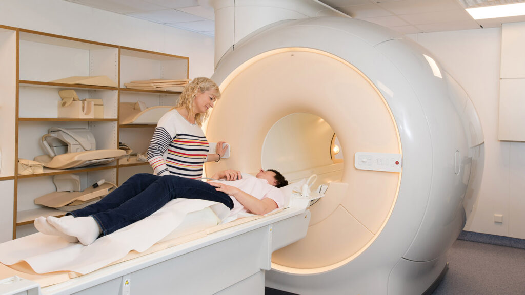
HALO MRI CENTER – 1720 Esplanade, Chico, CA 95926 – (530) 898-0500
Our team is MRI Accredited to provide you the best image quality possible.
At HALO MRI Center, you can expect:
- Accredited MRI Technicians
- Board-certified Radiologists
- Warm, compassionate, caring team members
- Same day / next day appointments
- Same day / next day image turnaround time
Vision: To provide readers with a comprehensive overview of the various types of MRI available, highlighting the advanced medical imaging techniques that are being used to diagnose and treat a wide range of medical conditions.
Inspiration: This blog is designed to be a valuable resource for healthcare professionals and patients alike, providing a deep dive into the world of MRI and the many different techniques and technologies that are available. By sharing insights and knowledge about the latest advances in MRI, we hope to inspire and inform readers, ultimately helping to improve patient outcomes and advance the field of medical imaging.
A Closer Look Inside: The Many Types of MRI Exams and Their Benefits
1. Conventional MRI: This is the most common type of MRI that uses a powerful magnet and radio waves to produce detailed images of the body’s internal structures. It can be used to detect a wide range of abnormalities in the body, including tumors, inflammation, and tissue damage.
2. Functional MRI (fMRI): This type of MRI is used to detect changes in blood flow in the brain, which can help to identify areas of the brain that are active during specific tasks or activities. It is often used to study brain function and to detect abnormalities such as tumors or brain injuries.
3. Magnetic Resonance Angiography (MRA): This type of MRI is used to produce detailed images of the body’s blood vessels, which can help to detect blockages, aneurysms, or other abnormalities. It is often used to diagnose heart disease, stroke, or other vascular conditions.
4. Diffusion-weighted imaging (DWI): This type of MRI is used to measure the movement of water molecules in the body’s tissues, which can help to identify areas of tissue damage or inflammation. It is often used to detect early signs of stroke, brain injuries, or tumors.
5. Magnetic Resonance Spectroscopy (MRS): This type of MRI is used to analyze the chemical composition of the body’s tissues, which can help to detect abnormalities in metabolism or cell function. It is often used to study brain function and to detect abnormalities such as tumors or neurological disorders.
6. Magnetic Resonance Elastography (MRE): This type of MRI is used to measure the stiffness of tissues, which can help to detect areas of tissue damage or disease. It is often used to detect liver disease or to monitor the progression of cancer.
7. Cardiac MRI: This type of MRI is used to produce detailed images of the heart and its structures, which can help to detect abnormalities such as heart disease or congenital heart defects. It is often used to diagnose and monitor heart disease and to plan cardiac surgery.
8. Breast MRI: This type of MRI is used to produce detailed images of the breast tissue, which can help to detect breast cancer or other abnormalities. It is often used to diagnose breast cancer in women with dense breast tissue or to monitor the effectiveness of breast cancer treatment.
9. Prostate MRI: This type of MRI is used to produce detailed images of the prostate gland, which can help to detect prostate cancer or other abnormalities. It is often used to diagnose prostate cancer and to guide prostate cancer treatment.
10. Pelvic MRI: This type of MRI is used to produce detailed images of the pelvic area, which can help to detect abnormalities in the reproductive or urinary systems. It is often used to diagnose and monitor gynecological or urological conditions.
11. Abdominal MRI: This type of MRI is used to produce detailed images of the abdominal area, which can help to detect abnormalities in the liver, pancreas, or other organs. It is often used to diagnose and monitor liver disease, pancreatic cancer, or other gastrointestinal conditions.
12. Head and Neck MRI: This type of MRI is used to produce detailed images of the head and neck, which can help to detect abnormalities in the brain, sinuses, or other structures. It is often used to diagnose and monitor brain tumors, neurological disorders, or sinus conditions.
13. Magnetic Resonance Angiography (MRA): This type of MRI is used to produce detailed images of the body’s blood vessels, which can help to detect blockages, aneurysms, or other abnormalities. It is often used to diagnose heart disease, stroke, or other vascular conditions.
14. Diffusion-weighted imaging (DWI): This type of MRI is used to measure the movement of water molecules in the body’s tissues, which can help to identify areas of tissue damage or inflammation. It is often used to detect early signs of stroke, brain injuries, or tumors.
15. Magnetic Resonance Spectroscopy (MRS): This type of MRI is used to analyze the chemical composition of the body’s tissues, which can help to detect abnormalities in metabolism or cell function. It is often used to study brain function and to detect abnormalities such as tumors or neurological disorders.
16. Extremity MRI: This type of MRI is used to produce detailed images of the arms or legs, which can help to detect abnormalities such as fractures or joint injuries. It is often used to diagnose and monitor conditions such as arthritis, bone tumors, or tendon or ligament injuries.
17. Whole-body MRI: This type of MRI is used to produce detailed images of the entire body, which can help to detect abnormalities such as tumors or other systemic diseases. It is often used for cancer screening or to monitor the progression of cancer or other diseases.
18. Perfusion MRI: This type of MRI is used to measure blood flow to different parts of the body, which can help to detect abnormalities such as stroke or tumor growth. It is often used to diagnose and monitor brain tumors, stroke, or other vascular conditions.
19. Susceptibility-weighted imaging (SWI): This type of MRI is used to produce highly detailed images of the brain and other structures, which can help to detect abnormalities such as small hemorrhages or iron deposits. It is often used to diagnose and monitor neurological conditions such as multiple sclerosis or Alzheimer’s disease.
20. Magnetic resonance neurography (MRN): This type of MRI is used to produce detailed images of nerves and surrounding structures, which can help to detect abnormalities such as nerve compression or injury. It is often used to diagnose and monitor conditions such as carpal tunnel syndrome, sciatica, or brachial plexus injuries.
21. MR arthrography: This type of MRI is used to produce detailed images of joints, which can help to detect abnormalities such as tears or inflammation. It is often used to diagnose and monitor conditions such as osteoarthritis, rotator cuff injuries, or cartilage damage.
22. MR enterography: This type of MRI is used to produce detailed images of the small intestine, which can help to diagnose and monitor conditions such as Crohn’s disease, ulcerative colitis, or small bowel tumors.
23. MR urography: This type of MRI is used to produce detailed images of the urinary tract, which can help to detect abnormalities such as kidney stones, tumors, or blockages. It is often used to diagnose and monitor conditions such as bladder or kidney cancer, or to evaluate the effectiveness of treatment for urinary tract conditions.
24. MR cholangiopancreatography (MRCP): This type of MRI is used to produce detailed images of the bile and pancreatic ducts, which can help to detect abnormalities such as blockages or tumors. It is often used to diagnose and monitor conditions such as pancreatic cancer, bile duct obstruction, or pancreatitis.
25. MR lymphography: This type of MRI is used to produce detailed images of the lymphatic system, which can help to detect abnormalities such as lymphedema or lymphoma. It is often used to diagnose and monitor the progression of cancer or other lymphatic system conditions.
26. MR colonography: This type of MRI is used to produce detailed images of the colon, which can help to detect abnormalities such as polyps or tumors. It is often used as an alternative to traditional colonoscopy, especially for patients who are unable to undergo a traditional colonoscopy.
27. MR venography: This type of MRI is used to produce detailed images of the veins, which can help to detect abnormalities such as blood clots or venous insufficiency. It is often used to diagnose and monitor conditions such as deep vein thrombosis, pulmonary embolism, or varicose veins.
28. MR angiography with contrast (MRA with contrast): This type of MRI uses a contrast agent to produce detailed images of blood vessels, which can help to detect abnormalities such as aneurysms or blood clots. It is often used to diagnose and monitor conditions such as peripheral artery disease or renal artery stenosis.
29. MR-guided focused ultrasound (MRgFUS): This type of MRI uses high-intensity focused ultrasound waves to heat and destroy tissue, which can be used to treat conditions such as uterine fibroids or tumors. It is a minimally invasive alternative to traditional surgery or radiation therapy.
30. MR spectroscopic imaging (MRSI): This type of MRI uses a specialized technique to measure the chemical composition of tissues, which can help to detect abnormalities such as tumors or metabolic disorders. It is often used to diagnose and monitor conditions such as brain tumors, prostate cancer, or multiple sclerosis.
31. MR perfusion-weighted imaging (PWI): This type of MRI uses a specialized technique to measure blood flow to tissues, which can help to detect abnormalities such as strokes or brain tumors. It is often used to diagnose and monitor neurological conditions, especially those affecting the brain.
32. MR diffusion tensor imaging (DTI): This type of MRI uses a specialized technique to measure the movement of water molecules in tissues, which can help to map the structure and connectivity of the brain’s white matter. It is often used to diagnose and monitor conditions such as traumatic brain injury, Alzheimer’s disease, or multiple sclerosis.
33. MR elastography of liver (MREL): This type of MRI is used to measure the stiffness of liver tissue, which can help to diagnose liver fibrosis and cirrhosis. It is a non-invasive alternative to traditional liver biopsies.
34. MR enteroclysis: This type of MRI is used to produce detailed images of the small intestine using a specialized technique that involves ingesting contrast material. It is often used to diagnose and monitor conditions such as Crohn’s disease or small bowel obstruction.
35. MR functional diffusion mapping (fDM): This type of MRI is used to measure changes in water diffusion in tissues over time, which can help to detect tumor growth and response to treatment. It is often used to monitor the progression of brain tumors.
36. MR thermography: This type of MRI is used to measure temperature changes in tissues, which can help to monitor the effectiveness of cancer treatment or to guide thermal ablation therapy for tumors.
37. MR cine imaging: This type of MRI produces images of moving organs, such as the heart, which can help to detect abnormalities such as valve defects or heart failure. It is often used to diagnose and monitor cardiac conditions.
38. MR neuroimaging with iron oxide nanoparticles: This type of MRI uses iron oxide nanoparticles to enhance contrast and image brain tissue. It is often used in research studies to investigate neurodegenerative diseases such as Parkinson’s and Alzheimer’s disease.
39. MR spectroscopy imaging of hyperpolarized 13C-labeled substrates: This type of MRI uses hyperpolarized 13C-labeled substrates to image metabolic processes in vivo. It is often used in research studies to investigate cancer metabolism.
40. MR angiography without contrast: This type of MRI uses specialized techniques such as time-of-flight (TOF) or phase-contrast (PC) to image blood vessels without the use of contrast material. It is often used in patients who are allergic to contrast material or who have impaired kidney function.
41. MR enteroclysis with anti-peristaltic agent: This type of MRI is similar to MR enteroclysis, but it uses an anti-peristaltic agent to temporarily stop intestinal contractions and improve image quality. It is often used to diagnose and monitor small bowel diseases such as Crohn’s disease or small bowel obstruction.
42. MR chemoembolization monitoring: This type of MRI is used to monitor the effectiveness of chemoembolization therapy for liver tumors. It involves imaging the liver before and after the procedure to evaluate tumor response and blood flow to the tumor.
43. MR-guided radiation therapy (MRgRT): This type of MRI is used to guide radiation therapy for cancer treatment. It uses real-time MRI imaging to track tumor motion and adjust the radiation beam accordingly, reducing radiation exposure to healthy tissue.
44. MR mammography (MRM): This type of MRI is used to screen for breast cancer in women who are at high risk or who have dense breast tissue. It produces highly detailed images of the breast tissue and can detect small tumors that may not be visible on mammography or ultrasound.
45. MR imaging of lung function: This type of MRI is used to image lung ventilation and perfusion, and can detect abnormalities such as lung disease or pulmonary embolism. It is often used as an alternative to traditional pulmonary function testing.
46. MR image-guided biopsy: This type of MRI is used to guide a biopsy needle to a specific location in the body for tissue sampling. It is often used to diagnose and monitor cancer or other conditions that cannot be diagnosed with imaging alone.
47. MR enterography with diffusion-weighted imaging (DWI): This type of MRI combines the specialized techniques of MR enterography and DWI to produce highly detailed images of the small intestine. It is often used to diagnose and monitor inflammatory bowel disease or small bowel tumors.
48. MR-guided cryoablation: This type of MRI is used to guide a cryoablation probe to a specific location in the body for tissue destruction. It is often used to treat tumors or other conditions that cannot be treated with surgery or radiation therapy.
49. MR neurography of the brachial plexus: This type of MRI is used to produce detailed images of the brachial plexus, which is a network of nerves that controls movement and sensation in the arm. It is often used to diagnose and monitor conditions such as thoracic outlet syndrome or brachial plexus injury.
Precision Imaging, Precision Medicine: The Role of MRI in Healthcare
50. MR spectroscopy imaging of lactate: This type of MRI is used to detect elevated levels of lactate in tissues, which can indicate the presence of tumors or other metabolic disorders. It is often used to diagnose and monitor cancer or other metabolic conditions.
51. MR urography with diffusion-weighted imaging (DWI): This type of MRI combines the specialized techniques of MR urography and DWI to produce highly detailed images of the urinary tract. It is often used to diagnose and monitor conditions such as kidney or bladder cancer.
52. MR imaging of cerebral blood flow (CBF): This type of MRI is used to measure blood flow to the brain, which can help to detect abnormalities such as stroke or Alzheimer’s disease. It is often used in research studies to investigate brain function and neurological disorders.
53. MR spectroscopy imaging of phosphorus-31 (31P): This type of MRI is used to image energy metabolism in tissues by measuring levels of phosphorus-containing compounds. It is often used in research studies to investigate metabolic disorders and neurodegenerative diseases.
54. MR angiography of the lower extremities: This type of MRI is used to produce detailed images of blood vessels in the legs and feet, which can help to diagnose and monitor conditions such as peripheral artery disease or deep vein thrombosis.
55. MR-guided laser interstitial thermal therapy (MRgLITT): This type of MRI is used to guide a laser probe to a specific location in the brain for thermal ablation of tumors or other conditions. It is often used as a minimally invasive alternative to traditional brain surgery or radiation therapy.
56. MR imaging of bone marrow: This type of MRI is used to produce detailed images of bone marrow, which can help to detect abnormalities such as bone marrow disorders or bone metastases. It is often used to diagnose and monitor conditions such as leukemia or multiple myeloma.
57. MR lymphangiography: This type of MRI is used to produce detailed images of the lymphatic system, which can help to detect abnormalities such as lymphedema or lymphatic malformations. It is often used to diagnose and monitor the progression of cancer or other lymphatic system conditions.
58. MR elastography of the brain: This type of MRI is used to measure the stiffness of brain tissue, which can help to detect abnormalities such as brain tumors or neurological disorders. It is often used in research studies to investigate brain function and disease.
59. MR elastography of the breast: This type of MRI is used to measure the stiffness of breast tissue, which can help to detect abnormalities such as breast cancer or fibroadenomas. It is often used in women with dense breast tissue or to monitor the effectiveness of breast cancer treatment.
60. MR-guided high-intensity focused ultrasound (MRgHIFU): This type of MRI is used to guide high-intensity focused ultrasound waves to a specific location in the body for thermal ablation of tumors or other conditions. It is often used as a minimally invasive alternative to traditional surgery or radiation therapy.
61. MR imaging of fat: This type of MRI is used to produce detailed images of fat tissue, which can help to detect abnormalities such as lipomas or fatty liver disease. It is often used to diagnose and monitor conditions such as obesity or metabolic disorders.
62. MR imaging of the temporomandibular joint (TMJ): This type of MRI is used to produce detailed images of the TMJ, which can help to diagnose and monitor conditions such as TMJ disorder or osteoarthritis of the jaw. It is often used in patients with chronic jaw pain or difficulty chewing.
63. MR-guided radiotherapy planning: This type of MRI is used to guide the planning of radiation therapy for cancer treatment. It uses MRI images to create a highly accurate 3D map of the tumor and surrounding tissues, which can improve the precision of radiation therapy and reduce the risk of damage to healthy tissues.
64. MR imaging of the peripheral nerves: This type of MRI is used to produce detailed images of the peripheral nerves, which can help to diagnose and monitor conditions such as nerve entrapment or neuropathy. It is often used in patients with chronic pain or numbness in the hands or feet.
65. MR spectroscopy imaging of sodium-23 (23Na): This type of MRI is used to image sodium ions in tissues, which can help to detect abnormalities such as changes in cell volume or tissue hydration. It is often used in research studies to investigate metabolic disorders and neurodegenerative diseases.
66. MR angiography of the carotid arteries: This type of MRI is used to produce detailed images of the carotid arteries in the neck, which can help to diagnose and monitor conditions such as carotid artery stenosis or atherosclerosis. It is often used in patients at high risk for stroke.
67. MR imaging of the liver with contrast: This type of MRI uses a contrast agent to produce detailed images of the liver, which can help to detect abnormalities such as liver tumors or cirrhosis. It is often used in patients who cannot undergo a computed tomography (CT) scan with contrast.
68. MR imaging of the spine with contrast: This type of MRI uses a contrast agent to produce detailed images of the spine, which can help to detect abnormalities such as herniated discs or spinal cord tumors. It is often used in patients with back pain or neurological symptoms.
69. MR-guided prostate biopsy: This type of MRI is used to guide a biopsy needle to a specific location in the prostate gland for tissue sampling. It is often used to diagnose and monitor prostate cancer or other conditions that cannot be diagnosed with imaging alone.
70. MR imaging of the eye: This type of MRI is used to produce detailed images of the eye and surrounding structures, which can help to diagnose and monitor conditions such as optic neuritis or retinal detachment. It is often used in patients with vision loss or eye pain.
71. MR imaging of the pituitary gland: This type of MRI is used to produce detailed images of the pituitary gland, which can help to detect abnormalities such as pituitary tumors or pituitary gland dysfunction. It is often used in patients with hormonal imbalances or neurological symptoms.
72. MR imaging of the temporomandibular joint (TMJ) with contrast: This type of MRI uses a contrast agent to produce highly detailed images of the TMJ, which can help to diagnose and monitor conditions such as TMJ disorder or osteoarthritis of the jaw. It is often used in patients with chronic jaw pain or difficulty chewing.
73. MR imaging of the placenta: This type of MRI is used to produce detailed images of the placenta, which can help to detect abnormalities such as placenta previa or placental insufficiency. It is often used in pregnant women with high-risk pregnancies or fetal growth problems.
74. MR imaging of the breast with contrast: This type of MRI uses a contrast agent to produce highly detailed images of the breast tissue, which can help to detect abnormalities such as breast cancer or suspicious lesions. It is often used in women with dense breast tissue or to monitor the effectiveness of breast cancer treatment.
75. MR elastography of the prostate: This type of MRI is used to measure the stiffness of prostate tissue, which can help to detect abnormalities such as prostate cancer or prostatitis. It is often used in men with elevated prostate-specific antigen (PSA) levels or other symptoms of prostate disease.
76. MR imaging of the ankle with contrast: This type of MRI uses a contrast agent to produce detailed images of the ankle joint, which can help to diagnose and monitor conditions such as arthritis or ankle sprains. It is often used in athletes or people with chronic ankle pain.
77. MR-guided transcranial magnetic stimulation (TMS): This type of MRI is used to guide TMS therapy for neurological conditions such as depression or chronic pain. It uses real-time MRI imaging to target specific areas of the brain with magnetic pulses, which can modulate brain activity and alleviate symptoms.
78. MR imaging of the kidney with contrast: This type of MRI uses a contrast agent to produce detailed images of the kidneys, which can help to detect abnormalities such as kidney tumors or cysts. It is often used in patients with chronic kidney disease or renal dysfunction.
79. MR imaging of the heart with contrast: This type of MRI uses a contrast agent to produce highly detailed images of the heart, which can help to diagnose and monitor conditions such as heart valve defects or heart failure. It is often used in patients with cardiovascular disease or at risk for heart disease.
80. MR-guided transurethral ultrasound ablation (TULSA): This type of MRI is used to guide a transurethral ultrasound probe to a specific location in the prostate gland for thermal ablation of tumors or other conditions. It is often used as a minimally invasive alternative to traditional surgery or radiation therapy for prostate cancer.
81. MR imaging of the pelvis with contrast: This type of MRI uses a contrast agent to produce detailed images of the pelvic region, which can help to diagnose and monitor conditions such as uterine fibroids or ovarian cysts. It is often used in women with gynecological problems or pelvic pain.
82. MR-guided breast biopsy: This type of MRI is used to guide a biopsy needle to a specific location in the breast for tissue sampling. It is often used to diagnose and monitor breast cancer or other conditions that cannot be diagnosed with imaging alone.
83. MR diffusion tensor imaging (DTI) of the brain: This type of MRI is used to measure the diffusion of water molecules in the brain, which can help to map the neural pathways that connect different regions of the brain. It is often used in research studies to investigate brain function and connectivity, and in clinical practice to evaluate neurological disorders.
84. MR imaging of the adrenal glands with contrast: This type of MRI uses a contrast agent to produce detailed images of the adrenal glands, which can help to detect abnormalities such as adrenal tumors or hyperplasia. It is often used in patients with adrenal insufficiency or other endocrine disorders.
85. MR imaging of the knee with diffusion-weighted imaging (DWI): This type of MRI combines the specialized techniques of MR imaging of the knee and DWI to produce highly detailed images of the knee joint, which can help to diagnose and monitor conditions such as meniscal tears or osteoarthritis.
86. MR imaging of the lung with hyperpolarized noble gases: This type of MRI uses hyperpolarized noble gases, such as helium or xenon, to produce images of lung function and ventilation. It is often used in research studies to investigate lung diseases such as asthma or chronic obstructive pulmonary disease (COPD).
87. MR spectroscopy imaging of fluorine-19 (19F): This type of MRI is used to image fluorine-containing compounds in tissues, which can help to detect abnormalities such as inflammation or infection. It is often used in research studies to investigate immune system function and inflammatory disorders.
88. MR imaging of the temporomandibular joint (TMJ) with open mouth imaging: This type of MRI uses specialized techniques to image the TMJ with the patient’s mouth in an open position, which can help to diagnose and monitor conditions such as TMJ dislocation or degenerative joint disease. It is often used in patients with chronic jaw pain or limited mouth opening.
89. MR imaging of the liver with gadoxetic acid: This type of MRI uses the contrast agent gadoxetic acid to produce highly detailed images of the liver, which can help to detect abnormalities such as liver cancer or cirrhosis. It is often used in patients with chronic liver disease or liver dysfunction.
90. MR imaging of the spleen with contrast: This type of MRI uses a contrast agent to produce detailed images of the spleen, which can help to detect abnormalities such as splenomegaly or splenic tumors. It is often used in patients with blood disorders or other splenic diseases.
91. MR-guided focused ultrasound (MRgFUS) thalamotomy: This type of MRI is used to guide a focused ultrasound beam to a specific location in the brain for thermal ablation of the thalamus, which can help to alleviate symptoms of neurological disorders such as tremors or chronic pain. It is often used as a minimally invasive alternative to traditional brain surgery.
92. MR imaging of the temporomandibular joint (TMJ) with dynamic imaging: This type of MRI uses specialized techniques to image the TMJ with dynamic movements of the jaw, which can help to diagnose and monitor conditions such as TMJ clicking or dislocation. It is often used in patients with chronic jaw pain or clicking sounds during jaw movements.
93. MR imaging of the pancreas with contrast: This type of MRI uses a contrast agent to produce detailed images of the pancreas, which can help to detect abnormalities such as pancreatic cancer or pancreatitis. It is often used in patients with pancreatic disease or at risk for pancreatic cancer.
94. MR perfusion imaging of the brain: This type of MRI is used to measure blood flow to the brain, which can help to detect abnormalities such as stroke or brain tumors. It is often used in clinical practice to evaluate patients with acute stroke symptoms or in research studies to investigate brain function and disease.
95. MR imaging of the temporomandibular joint (TMJ) with cine imaging: This type of MRI uses specialized techniques to image the TMJ with dynamic movements of the jaw in real time, which can help to diagnose and monitor conditions such as TMJ clicking or subluxation. It is often used in patients with chronic jaw pain or clicking sounds during jaw movements.
96. MR imaging of the biliary tree with contrast: This type of MRI uses a contrast agent to produce detailed images of the biliary tree, which can help to detect abnormalities such as biliary strictures or bile duct tumors. It is often used in patients with biliary disease or at risk for liver cancer.
97. MR spectroscopy imaging of carbon-13 (13C): This type of MRI is used to image metabolic activity in tissues by measuring levels of carbon-13-containing compounds. It is often used in research studies to investigate metabolic disorders and neurodegenerative diseases.
98. MR imaging of the prostate with diffusion-weighted imaging (DWI): This type of MRI combines the specialized techniques of MR imaging of the prostate and DWI to produce highly detailed images of the prostate gland, which can help to detect abnormalities such as prostate cancer or prostatitis. It is often used in men with elevated prostate-specific antigen (PSA) levels or other symptoms of prostate disease.
99. MR imaging of the brainstem: This type of MRI is used to produce detailed images of the brainstem, which controls many vital functions such as breathing, heart rate, and blood pressure. It is often used in patients with neurological disorders or traumatic brain injury.
Don’t Miss a Thing: The Comprehensive Guide to MRI and Its Medical Applications
100. MR imaging of the fetal brain: This type of MRI is used to produce detailed images of the developing fetal brain, which can help to detect abnormalities such as neural tube defects or brain malformations. It is often used in pregnant women with high-risk pregnancies or fetal growth problems.
101. MR imaging of the placenta with diffusion-weighted imaging (DWI): This type of MRI combines the specialized techniques of MR imaging of the placenta and DWI to produce highly detailed images of the placenta, which can help to detect abnormalities such as placental insufficiency or intrauterine growth restriction. It is often used in pregnant women with high-risk pregnancies or fetal growth problems.
102. MR imaging of the head and neck with contrast: This type of MRI uses a contrast agent to produce highly detailed images of the head and neck, which can help to detect abnormalities such as head and neck tumors or vascular malformations. It is often used in patients with head and neck cancer or vascular disorders.
103. MR elastography of the liver: This type of MRI is used to measure the stiffness of liver tissue, which can help to detect abnormalities such as liver fibrosis or cirrhosis. It is often used in patients with chronic liver disease or liver dysfunction.
104. MR imaging of the spinal cord: This type of MRI is used to produce detailed images of the spinal cord, which can help to detect abnormalities such as spinal cord tumors or multiple sclerosis. It is often used in patients with neurological symptoms or traumatic spinal cord injury.
105. MR imaging of the breast with diffusion-weighted imaging (DWI): This type of MRI combines the specialized techniques of MR imaging of the breast and DWI to produce highly detailed images of the breast tissue, which can help to detect abnormalities such as breast cancer or suspicious lesions.
106. MR imaging of the knee with proton density-weighted imaging (PDWI): This type of MRI combines the specialized techniques of MR imaging of the knee and PDWI to produce highly detailed images of the knee joint, which can help to diagnose and monitor conditions such as meniscal tears or osteoarthritis.
107. MR imaging of the ear with contrast: This type of MRI uses a contrast agent to produce detailed images of the ear and surrounding structures, which can help to detect abnormalities such as inner ear disorders or acoustic neuromas. It is often used in patients with hearing loss or balance problems.
108. MR imaging of the wrist with contrast: This type of MRI uses a contrast agent to produce detailed images of the wrist joint, which can help to diagnose and monitor conditions such as wrist arthritis or carpal tunnel syndrome. It is often used in patients with chronic wrist pain or swelling.
109. MR imaging of the ankle with proton density-weighted imaging (PDWI): This type of MRI combines the specialized techniques of MR imaging of the ankle and PDWI to produce highly detailed images of the ankle joint, which can help to diagnose and monitor conditions such as ankle arthritis or ligament tears.
110. MR imaging of the adrenal glands with MR spectroscopy: This type of MRI combines the specialized techniques of MR imaging of the adrenal glands and MR spectroscopy to produce highly detailed images of adrenal function and metabolism. It is often used in patients with adrenal disorders or hormonal imbalances.
111. MR-guided high-intensity focused ultrasound (MR-HIFU): This type of MRI is used to guide a focused ultrasound beam to a specific location in the body for thermal ablation of tumors or other conditions. It is often used as a minimally invasive alternative to traditional surgery or radiation therapy for cancer treatment.
112. MR imaging of the shoulder with diffusion-weighted imaging (DWI): This type of MRI combines the specialized techniques of MR imaging of the shoulder and DWI to produce highly detailed images of the shoulder joint, which can help to diagnose and monitor conditions such as rotator cuff tears or shoulder impingement syndrome.
113. MR imaging of the liver with diffusion-weighted imaging (DWI): This type of MRI combines the specialized techniques of MR imaging of the liver and DWI to produce highly detailed images of the liver, which can help to detect abnormalities such as liver cancer or cirrhosis.
114. MR imaging of the pelvis with diffusion-weighted imaging (DWI): This type of MRI combines the specialized techniques of MR imaging of the pelvis and DWI to produce highly detailed images of the pelvic region, which can help to diagnose and monitor conditions such as pelvic inflammatory disease or gynecological tumors.
115. MR imaging of the chest with contrast: This type of MRI uses a contrast agent to produce detailed images of the chest, which can help to detect abnormalities such as lung cancer or pulmonary embolism. It is often used in patients with respiratory symptoms or at risk for lung disease.
116. MR imaging of the cervical spine with contrast: This type of MRI uses a contrast agent to produce detailed images of the cervical spine, which can help to detect abnormalities such as spinal cord compression or herniated discs. It is often used in patients with neck pain or neurological symptoms.
117. MR imaging of the elbow with proton density-weighted imaging (PDWI): This type of MRI combines the specialized techniques of MR imaging of the elbow and PDWI to produce highly detailed images of the elbow joint, which can help to diagnose and monitor conditions such as tennis elbow or elbow arthritis.
118. MR imaging of the foot with proton density-weighted imaging (PDWI): This type of MRI combines the specialized techniques of MR imaging of the foot and PDWI to produce highly detailed images of the foot and ankle joints, which can help to diagnose and monitor conditions such as plantar fasciitis or ankle fractures.
119. MR imaging of the spine with diffusion tensor imaging (DTI): This type of MRI combines the specialized techniques of MR imaging of the spine and DTI to produce highly detailed images of the spinal cord and nerve pathways, which can help to diagnose and monitor conditions such as spinal cord injury or multiple sclerosis.
120. MR imaging of the eye with diffusion tensor imaging (DTI): This type of MRI combines the specialized techniques of MR imaging of the eye and DTI to produce highly detailed images of the optic nerve and visual pathways, which can help to diagnose and monitor conditions such as glaucoma or optic neuritis.
121. MR imaging of the heart with T1 mapping: This type of MRI uses specialized techniques to measure the T1 relaxation time of the heart tissue, which can help to detect abnormalities such as heart failure or myocardial infarction. It is often used in patients with cardiovascular disease or at risk for heart disease.
122. MR imaging of the spinal cord with dynamic contrast enhancement (DCE): This type of MRI combines the specialized techniques of MR imaging of the spine and DCE to produce highly detailed images of blood flow and permeability in the spinal cord, which can help to detect abnormalities such as spinal cord tumors or inflammation.
123. MR imaging of the wrist with diffusion tensor imaging (DTI): This type of MRI combines the specialized techniques of MR imaging of the wrist and DTI to produce highly detailed images of the wrist joint, which can help to diagnose and monitor conditions such as carpal tunnel syndrome or wrist sprains.
124. MR imaging of the liver with hepatocyte-specific contrast agents: This type of MRI uses specialized contrast agents that are taken up by liver cells to produce highly detailed images of liver function and metabolism. It is often used in patients with liver disease or liver dysfunction.
125. MR imaging of the neck with diffusion tensor imaging (DTI): This type of MRI uses specialized techniques to measure the diffusion of water molecules in neck tissue in multiple directions, which can help to detect abnormalities such as cervical spinal cord injuries or tumors.
126. MR imaging of the brain with arterial spin labeling (ASL): This type of MRI uses specialized techniques to measure blood flow to the brain, which can help to detect abnormalities such as stroke or brain tumors. It is often used in research studies to investigate brain function and disease.
127. MR imaging of the uterus with contrast: This type of MRI uses a contrast agent to produce detailed images of the uterus, which can help to detect abnormalities such as fibroids or endometrial cancer. It is often used in patients with gynecological symptoms or at risk for uterine disease.
128. MR imaging of the wrist with contrast: This type of MRI uses a contrast agent to produce highly detailed images of the wrist joint, which can help to diagnose and monitor conditions such as wrist arthritis or carpal tunnel syndrome. It is often used in patients with chronic wrist pain or swelling.
129. MR imaging of the breast with contrast-enhanced spectroscopy: This type of MRI combines the specialized techniques of MR imaging of the breast and MR spectroscopy to produce highly detailed images of breast tissue metabolism and function. It is often used in patients with breast cancer or at risk for breast cancer.
130. MR imaging of the abdomen and pelvis with diffusion-weighted imaging (DWI): This type of MRI combines the specialized techniques of MR imaging of the abdomen and pelvis and DWI to produce highly detailed images of the abdominal and pelvic regions, which can help to diagnose and monitor conditions such as inflammatory bowel disease or pelvic tumors.
131. MR imaging of the ankle with contrast: This type of MRI uses a contrast agent to produce highly detailed images of the ankle joint, which can help to diagnose and monitor conditions such as ankle arthritis or ligament tears.
132. MR imaging of the head and neck with proton density-weighted imaging (PDWI): This type of MRI combines the specialized techniques of MR imaging of the head and neck and PDWI to produce highly detailed images of the skull and cervical spine, which can help to diagnose and monitor conditions such as skull fractures or cervical spinal cord injury.
133. MR imaging of the abdomen and pelvis with dynamic contrast-enhanced imaging (DCE): This type of MRI combines the specialized techniques of MR imaging of the abdomen and pelvis and DCE to produce highly detailed images of blood flow and permeability in the abdominal and pelvic regions, which can help to diagnose and monitor conditions such as liver cancer or pelvic inflammatory disease.
134. MR imaging of the pancreas with diffusion-weighted imaging (DWI): This type of MRI combines the specialized techniques of MR imaging of the pancreas and DWI to produce highly detailed images of the pancreas, which can help to detect abnormalities such as pancreatic cancer or pancreatitis.
135. MR imaging of the spine with T2 mapping: This type of MRI uses specialized techniques to measure the T2 relaxation time of the spinal cord and nerve roots, which can help to detect abnormalities such as spinal cord injury or herniated discs.
136. MR imaging of the kidney with contrast: This type of MRI uses a contrast agent to produce detailed images of the kidney, which can help to detect abnormalities such as kidney stones or renal tumors. It is often used in patients with renal disease or at risk for kidney disease.
137. MR imaging of the ankle with diffusion tensor imaging (DTI): This type of MRI combines the specialized techniques of MR imaging of the ankle and DTI to produce highly detailed images of the ankle joint, which can help to diagnose and monitor conditions such as ankle ligament tears or instability.
138. MR imaging of the prostate with MR spectroscopy: This type of MRI combines the specialized techniques of MR imaging of the prostate with spectroscopy to measure the chemical composition of prostate tissue. By analyzing the chemical signals in the tissue, MR spectroscopy can help to distinguish between healthy prostate tissue and cancerous tissue. It is often used to guide biopsies of the prostate or to monitor the progression of prostate cancer.
139. MR imaging of the brain with resting-state functional MRI (rs-fMRI): This type of MRI is used to investigate the functional connectivity of different regions of the brain by measuring fluctuations in blood oxygen level-dependent (BOLD) signals. It is often used in research studies to investigate brain function and disease.
140. MR imaging of the pancreas with secretin stimulation: This type of MRI uses a hormone called secretin to stimulate pancreatic function and produce detailed images of the pancreas, which can help to detect abnormalities such as pancreatic tumors or chronic pancreatitis. It is often used in patients with pancreatic disease or at risk for pancreatic cancer.
141. MR imaging of the hip with contrast: This type of MRI uses a contrast agent to produce highly detailed images of the hip joint, which can help to diagnose and monitor conditions such as hip arthritis or labral tears.
142. MR imaging of the foot with contrast: This type of MRI uses a contrast agent to produce highly detailed images of the foot and ankle joints, which can help to diagnose and monitor conditions such as plantar fasciitis or ankle fractures.
143. MR imaging of the neck with proton density-weighted imaging (PDWI): This type of MRI combines the specialized techniques of MR imaging of the neck and PDWI to produce highly detailed images of the cervical spine and neck muscles, which can help to diagnose and monitor conditions such as cervical radiculopathy or neck strain.
144. MR imaging of the liver with T1 mapping: This type of MRI uses specialized techniques to measure the T1 relaxation time of liver tissue, which can help to detect abnormalities such as liver fibrosis or cirrhosis. It is often used in patients with chronic liver disease or liver dysfunction.
145. MR imaging of the spinal cord with T2* mapping: This type of MRI uses specialized techniques to measure the T2* relaxation time of the spinal cord, which can help to detect abnormalities such as spinal cord hemorrhage or demyelination.
146. MR imaging of the prostate with diffusion tensor imaging (DTI): This type of MRI combines the specialized techniques of MR imaging of the prostate and DTI to produce highly detailed images of the prostate gland and adjacent tissues, which can help to detect abnormalities such as prostate cancer or prostatitis.
147. MR imaging of the foot with diffusion-weighted imaging (DWI): This type of MRI combines the specialized techniques of MR imaging of the foot and DWI to produce highly detailed images of the foot and ankle joints, which can help to diagnose and monitor conditions such as gout or rheumatoid arthritis.
148. MR imaging of the liver with diffusion tensor imaging (DTI): This type of MRI combines the specialized techniques of MR imaging of the liver and DTI to produce highly detailed images of liver tissue microstructure and function, which can help to detect abnormalities such as liver fibrosis or cirrhosis.
149. MR imaging of the pelvis with contrast-enhanced spectroscopy: This type of MRI combines the specialized techniques of MR imaging of the pelvis and MR spectroscopy to produce highly detailed images of pelvic tissue metabolism and function. It is often used in patients with gynecological or urological conditions.
Empowering Healthcare Professionals: The Benefits of Advanced MRI Technology
150. MR imaging of the brain with susceptibility-weighted imaging (SWI): This type of MRI uses specialized techniques to produce highly detailed images of the brain’s venous vasculature, which can help to diagnose and monitor conditions such as cerebral venous thrombosis or arteriovenous malformations.
151. MR imaging of the heart with cine imaging: This type of MRI uses specialized techniques to produce high-resolution images of the beating heart, which can help to detect abnormalities such as heart failure or valvular disease. It is often used in patients with cardiovascular disease or at risk for heart disease.
152. MR imaging of the prostate with dynamic contrast-enhanced imaging (DCE): This type of MRI combines the specialized techniques of MR imaging of the prostate and DCE to produce highly detailed images of blood flow and permeability in the prostate gland, which can help to detect abnormalities such as prostate cancer or benign prostatic hyperplasia (BPH).
153. MR imaging of the knee with magnetization transfer imaging (MTI): This type of MRI uses specialized techniques to measure the magnetization transfer between water molecules and macromolecules in the knee joint, which can help to detect abnormalities such as cartilage degradation or meniscal injury.
154. MR imaging of the chest with cardiac-gated imaging: This type of MRI uses specialized techniques to synchronize image acquisition with the cardiac cycle, which can help to produce high-quality images of the heart and great vessels. It is often used in patients with congenital heart disease or at risk for cardiac dysfunction.
155. MR imaging of the brain with perfusion-weighted imaging (PWI): This type of MRI uses specialized techniques to measure cerebral blood flow and volume, which can help to diagnose and monitor conditions such as stroke or brain tumors.
156. MR imaging of the liver with hepatobiliary contrast agents: This type of MRI uses specialized contrast agents that are taken up by liver cells and excreted into the bile, which can help to detect abnormalities such as liver cancer or liver disease.
157. MR imaging of the breast with quantitative T2 mapping: This type of MRI uses specialized techniques to measure the T2 relaxation time of breast tissue, which can help to detect abnormalities such as breast cancer or fibroadenomas.
158. MR imaging of the foot with magnetic resonance neurography (MRN): This type of MRI uses specialized techniques to produce highly detailed images of peripheral nerves in the foot and ankle, which can help to diagnose and monitor conditions such as peripheral neuropathy or tarsal tunnel syndrome.
159. MR imaging of the brain with diffusion kurtosis imaging (DKI): This type of MRI uses specialized techniques to measure the diffusion kurtosis of water molecules in the brain, which can help to detect abnormalities such as traumatic brain injury or brain tumors.
160. MR imaging of the lung with oxygen-enhanced imaging: This type of MRI uses specialized techniques to measure the oxygen content of lung tissue, which can help to detect abnormalities such as pulmonary fibrosis or lung cancer.
161. MR imaging of the breast with diffusion-weighted imaging (DWI): This type of MRI uses specialized techniques to measure the diffusion of water molecules in breast tissue, which can help to detect abnormalities such as breast cancer or fibroadenomas.
162. MR imaging of the liver with magnetic resonance elastography (MRE): This type of MRI uses specialized techniques to measure the stiffness of liver tissue, which can help to detect abnormalities such as liver fibrosis or cirrhosis.
163. MR imaging of the brain with susceptibility tensor imaging (STI): This type of MRI uses specialized techniques to measure the anisotropy of magnetic susceptibility in the brain, which can help to detect abnormalities such as multiple sclerosis or cerebral microbleeds.
164. MR imaging of the knee with diffusion tensor imaging (DTI): This type of MRI combines the specialized techniques of MR imaging of the knee and DTI to produce highly detailed images of the knee joint, which can help to diagnose and monitor conditions such as knee ligament tears or cartilage degeneration.
165. MR imaging of the neck with magnetic resonance angiography (MRA): This type of MRI uses specialized techniques to produce highly detailed images of the blood vessels in the neck, which can help to detect abnormalities such as carotid artery stenosis or vertebral artery dissection.
166. MR imaging of the pancreas with magnetic resonance cholangiopancreatography (MRCP): This type of MRI uses specialized techniques to produce highly detailed images of the pancreatic and biliary ducts, which can help to detect abnormalities such as pancreaticobiliary cancers or gallstones.
167. MR imaging of the liver with dual-energy imaging: This type of MRI uses specialized techniques to measure the energy of X-ray photons absorbed by liver tissue, which can help to detect abnormalities such as liver tumors or fatty liver disease.
168. MR imaging of the spine with spinal cord fMRI: This type of MRI uses specialized techniques to measure the functional connectivity of the spinal cord and brain, which can help to detect abnormalities such as spinal cord injury or degenerative disc disease.
169. MR imaging of the liver with arterial spin labeling (ASL): This type of MRI uses specialized techniques to measure the blood flow to liver tissue, which can help to detect abnormalities such as liver tumors or cirrhosis.
170. MR imaging of the chest with diffusion-weighted imaging (DWI): This type of MRI combines the specialized techniques of MR imaging of the chest and DWI to produce highly detailed images of the lung and mediastinum, which can help to detect abnormalities such as lung cancer or lymphoma.
171. MR imaging of the liver with delayed hepatobiliary phase imaging: This type of MRI uses specialized techniques to produce highly detailed images of the liver after the administration of a hepatobiliary contrast agent, which can help to detect abnormalities such as liver tumors or bile duct obstruction.
172. MR imaging of the brain with susceptibility contrast imaging (SCI): This type of MRI uses specialized techniques to produce highly detailed images of iron accumulation in the brain, which can help to detect abnormalities such as Parkinson’s disease or Alzheimer’s disease.
173. MR imaging of the knee with magnetic resonance spectroscopic imaging (MRSI): This type of MRI uses specialized techniques to measure the chemical composition of knee tissue, which can help to detect abnormalities such as osteoarthritis or cartilage damage.
174. MR imaging of the brain with fluid-attenuated inversion recovery (FLAIR): This type of MRI uses specialized techniques to suppress the signal from cerebrospinal fluid and produce highly detailed images of brain tissue, which can help to detect abnormalities such as multiple sclerosis or brain tumors.
175. MR imaging of the spine with MR myelography: This type of MRI uses specialized techniques to produce highly detailed images of the spinal canal and nerve roots, which can help to detect abnormalities such as spinal stenosis or herniated discs.
176. MR imaging of the liver with diffusion kurtosis imaging (DKI): This type of MRI combines the specialized techniques of MR imaging of the liver and DKI to produce highly detailed images of liver tissue microstructure and function, which can help to detect abnormalities such as liver fibrosis or cirrhosis.
177. MR imaging of the brain with time-of-flight (TOF) angiography: This type of MRI uses specialized techniques to produce highly detailed images of the blood vessels in the brain, which can help to detect abnormalities such as aneurysms or arteriovenous malformations.
178. MR imaging of the breast with breast-specific gamma imaging (BSGI): This type of MRI uses a radioactive tracer to produce highly detailed images of breast tissue, which can help to detect abnormalities such as breast cancer or benign breast lesions.
179. MR imaging of the liver with hepatocyte-specific contrast agents: This type of MRI uses specialized contrast agents that are taken up by liver cells and produce highly detailed images of liver tissue, which can help to detect abnormalities such as liver tumors or cirrhosis.
180. MR imaging of the brain with arterial spin labeling (ASL) perfusion: This type of MRI uses specialized techniques to measure cerebral blood flow in the brain, which can help to detect abnormalities such as stroke or Alzheimer’s disease.
181. MR imaging of the knee with magnetic resonance fingerprinting (MRF): This type of MRI uses specialized techniques to measure multiple tissue properties simultaneously, which can help to detect abnormalities such as cartilage damage or ligament tears.
182. MR imaging of the chest with proton radiography: This type of MRI uses specialized techniques to measure the proton density of lung tissue, which can help to detect abnormalities such as lung cancer or pulmonary fibrosis.
183. MR imaging of the spine with magnetic resonance neurography (MRN): This type of MRI uses specialized techniques to produce highly detailed images of peripheral nerves in the spinal canal, which can help to diagnose and monitor conditions such as spinal cord injury or lumbar radiculopathy.
184. MR imaging of the brain with magnetic resonance elastography (MRE): This type of MRI uses specialized techniques to measure the stiffness of brain tissue, which can help to detect abnormalities such as traumatic brain injury or brain tumors.
185. MR imaging of the abdomen with balanced steady-state free precession (bSSFP): This type of MRI uses specialized techniques to produce highly detailed images of abdominal organs and structures, which can help to diagnose and monitor conditions such as abdominal aortic aneurysms or inflammatory bowel disease.
186. MR imaging of the heart with diffusion tensor imaging (DTI): This type of MRI uses specialized techniques to measure the diffusion of water molecules in the heart, which can help to detect abnormalities such as cardiomyopathy or heart failure.
187. MR imaging of the brain with T2 relaxometry: This type of MRI uses specialized techniques to measure the T2 relaxation time of brain tissue, which can help to detect abnormalities such as cerebral edema or demyelination.
188. MR imaging of the chest with magnetic resonance ventilation imaging (MRVI): This type of MRI uses specialized techniques to produce highly detailed images of lung ventilation, which can help to diagnose and monitor conditions such as chronic obstructive pulmonary disease (COPD) or asthma.
189. MR imaging of the heart with phase-contrast imaging: This type of MRI uses specialized techniques to measure the flow of blood in the heart and great vessels, which can help to detect abnormalities such as valvular disease or congenital heart defects.
190. MR imaging of the brain with magnetic resonance spectroscopy (MRS): This type of MRI uses specialized techniques to measure the chemical composition of brain tissue, which can help to detect abnormalities such as brain tumors or Alzheimer’s disease.
191. MR imaging of the abdomen with magnetic resonance enterography (MRE): This type of MRI uses specialized techniques to produce highly detailed images of the small intestine, which can help to diagnose and monitor conditions such as Crohn’s disease or small bowel obstruction.
192. MR imaging of the hip with magnetic resonance arthrography (MRA): This type of MRI uses specialized techniques to produce highly detailed images of the hip joint, which can help to diagnose and monitor conditions such as hip labral tears or avascular necrosis.
193. MR imaging of the knee with magnetization transfer contrast imaging (MTCI): This type of MRI uses specialized techniques to measure the magnetization transfer between water molecules and macromolecules in the knee joint, which can help to detect abnormalities such as cartilage damage or osteoarthritis.
194. MR imaging of the brain with magnetization transfer ratio (MTR) imaging: This type of MRI uses specialized techniques to measure the magnetization transfer between water molecules and macromolecules in brain tissue, which can help to detect abnormalities such as multiple sclerosis or cerebral atrophy.
195. MR imaging of the liver with diffusion-weighted imaging (DWI): This type of MRI uses specialized techniques to measure the diffusion of water molecules in liver tissue, which can help to detect abnormalities such as liver tumors or cirrhosis.
196. MR imaging of the abdomen with magnetic resonance elastography (MRE): This type of MRI uses specialized techniques to measure the stiffness of abdominal organs and tissues, which can help to detect abnormalities such as liver fibrosis or pancreatic cancer.
197. MR imaging of the brain with quantitative susceptibility mapping (QSM): This type of MRI uses specialized techniques to measure the magnetic susceptibility of brain tissue, which can help to detect abnormalities such as cerebral microbleeds or iron accumulation.
198. MR imaging of the brain with 3D time-of-flight (TOF) angiography: This type of MRI uses specialized techniques to produce highly detailed 3D images of the blood vessels in the brain, which can help to detect abnormalities such as aneurysms or arteriovenous malformations.
199. MR imaging of the spine with spinal cord tractography: This type of MRI uses specialized techniques to produce highly detailed images of the spinal cord and nerve tracts, which can help to detect abnormalities such as spinal cord injury or multiple sclerosis.
The Cutting-Edge of Medical Diagnosis: Discovering the Many Types of MRI Exams
200. MR imaging of the knee with multi-echo spin-echo (MESE) imaging: This type of MRI uses specialized techniques to produce highly detailed images of the knee joint, which can help to detect abnormalities such as ligament tears or osteochondritis dissecans.
201. MR imaging of the heart with cardiac magnetic resonance spectroscopy (CMRS): This type of MRI uses specialized techniques to measure the metabolic activity of heart tissue, which can help to detect abnormalities such as myocardial infarction or heart failure.MR imaging of the chest with magnetic resonance fingerprinting (MRF): This type of MRI uses specialized techniques to measure multiple tissue properties simultaneously in the chest, which can help to detect abnormalities such as lung cancer or pulmonary fibrosis.
202. MR imaging of the brain with arterial spin labeling (ASL) fMRI: This type of MRI uses specialized techniques to measure the blood flow to the brain during functional tasks, which can help to detect abnormalities such as epilepsy or stroke.
203. MR imaging of the liver with blood oxygen level-dependent (BOLD) imaging: This type of MRI uses specialized techniques to measure the changes in oxygen levels in liver tissue during functional tasks, which can help to detect abnormalities such as liver tumors or liver cirrhosis.
204. MR imaging of the brain with proton magnetic resonance spectroscopy (1H MRS): This type of MRI uses specialized techniques to measure the levels of brain metabolites, which can help to detect abnormalities such as brain tumors or metabolic disorders.
205. MR imaging of the pelvis with magnetic resonance urography (MRU): This type of MRI uses specialized techniques to produce highly detailed images of the urinary tract, which can help to detect abnormalities such as kidney stones or bladder tumors.
206. MR imaging of the prostate with diffusion-weighted imaging (DWI): This type of MRI uses specialized techniques to measure the diffusion of water molecules in prostate tissue, which can help to detect abnormalities such as prostate cancer or prostatitis.
207. MR imaging of the brain with susceptibility-weighted imaging (SWI): This type of MRI uses specialized techniques to produce highly detailed images of iron and other mineral deposits in brain tissue, which can help to detect abnormalities such as cerebral microbleeds or neurodegenerative diseases.
208. MR imaging of the liver with gadoxetate disodium-enhanced imaging: This type of MRI uses a contrast agent that is taken up by liver cells to produce highly detailed images of liver tissue, which can help to detect abnormalities such as liver tumors or cirrhosis.
209. MR imaging of the knee with T2 mapping: This type of MRI uses specialized techniques to measure the T2 relaxation time of knee tissue, which can help to detect abnormalities such as cartilage damage or osteoarthritis.
210. MR imaging of the spine with diffusion tensor tractography (DTT): This type of MRI uses specialized techniques to produce highly detailed images of the spinal cord and nerve tracts, which can help to detect abnormalities such as spinal cord injury or multiple sclerosis.
211. MR imaging of the brain with functional connectivity magnetic resonance imaging (fcMRI): This type of MRI uses specialized techniques to measure the functional connectivity of different brain regions, which can help to detect abnormalities such as Alzheimer’s disease or depression.
212. MR imaging of the chest with ultra-short echo time (UTE) imaging: This type of MRI uses specialized techniques to produce highly detailed images of lung tissue with very short echo times, which can help to detect abnormalities such as lung nodules or emphysema.
213. MR imaging of the liver with gadoxetic acid-enhanced imaging: This type of MRI uses a contrast agent that is taken up by liver cells to produce highly detailed images of liver tissue, which can help to detect abnormalities such as liver tumors or cirrhosis.
214. MR imaging of the brain with dynamic susceptibility contrast (DSC) perfusion: This type of
215. MR imaging of the breast with diffusion-weighted imaging (DWI): This type of MRI uses specialized techniques to measure the diffusion of water molecules in breast tissue, which can help to detect abnormalities such as breast cancer or benign breast lesions.
216. MR imaging of the brain with susceptibility-weighted perfusion imaging (SWPI): This type of MRI uses specialized techniques to measure the magnetic susceptibility of blood in brain tissue, which can help to detect abnormalities such as stroke or cerebral microbleeds.
217. MR imaging of the liver with hepatic arterial phase imaging (HAP): This type of MRI uses specialized techniques to produce highly detailed images of the blood vessels in the liver during the arterial phase of contrast agent injection, which can help to detect abnormalities such as liver tumors or cirrhosis.
218. MR imaging of the brain with high-resolution functional MRI (hr-fMRI): This type of MRI uses specialized techniques to produce highly detailed images of brain function, which can help to detect abnormalities such as epilepsy or Alzheimer’s disease.
219. MR imaging of the prostate with T2*-weighted imaging: This type of MRI uses specialized techniques to produce highly detailed images of prostate tissue, which can help to detect abnormalities such as prostate cancer or prostatitis.
220. MR imaging of the brain with blood oxygen level-dependent (BOLD) fMRI: This type of MRI uses specialized techniques to measure the changes in oxygen levels in brain tissue during functional tasks, which can help to detect abnormalities such as epilepsy or Alzheimer’s disease.
221. MR imaging of the chest with magnetic resonance lymphography (MRL): This type of MRI uses specialized techniques to produce highly detailed images of the lymphatic system in the chest, which can help to detect abnormalities such as lymphedema or lymphoma.
222. MR imaging of the brain with high angular resolution diffusion imaging (HARDI): This type of MRI uses specialized techniques to measure the diffusion of water molecules in brain tissue in multiple directions, which can help to detect abnormalities such as white matter damage or multiple sclerosis.
223. MR imaging of the spine with gadofosveset-enhanced imaging: This type of MRI uses a contrast agent that is taken up by the spinal cord and nerve roots to produce highly detailed images of the spinal canal, which can help to detect abnormalities such as spinal cord tumors or herniated discs.
224. MR imaging of the knee with sodium magnetic resonance imaging (23Na-MRI): This type of MRI uses specialized techniques to measure the concentration of sodium ions in knee tissue, which can help to detect abnormalities such as osteoarthritis or cartilage damage.
225. MR imaging of the heart with tagged MRI: This type of MRI uses specialized techniques to produce highly detailed images of the motion of heart tissue, which can help to detect abnormalities such as heart failure or myocardial infarction.
226. MR imaging of the brain with functional diffusion mapping (fDM): This type of MRI uses specialized techniques to measure changes in water diffusion in brain tissue during functional tasks, which can help to detect abnormalities such as brain tumors or ischemic stroke.
227. MR imaging of the breast with breast magnetic resonance imaging-guided biopsy (MRI-guided biopsy): This type of MRI uses specialized techniques to guide a biopsy needle to a specific location in the breast, which can help to diagnose breast cancer or benign breast lesions.
228. MR imaging of the liver with T1 mapping: This type of MRI uses specialized techniques to measure the T1 relaxation time of liver tissue, which can help to detect abnormalities such as liver fibrosis or cirrhosis.
229. MR imaging of the brain with MR elastography (MRE): This type of MRI
230. MR imaging of the brain with magnetic resonance encephalography (MREG): This type of MRI uses specialized techniques to measure the dynamic changes in brain activity and blood flow, which can help to detect abnormalities such as epilepsy or brain tumors.
231. MR imaging of the liver with arterial spin labeling (ASL) perfusion imaging: This type of MRI uses specialized techniques to measure the blood flow to the liver, which can help to detect abnormalities such as liver tumors or cirrhosis.
232. MR imaging of the chest with magnetic resonance angiography (MRA): This type of MRI uses specialized techniques to produce highly detailed images of the blood vessels in the chest, which can help to detect abnormalities such as aneurysms or pulmonary embolism.
233. MR imaging of the spine with T1-weighted imaging: This type of MRI uses specialized techniques to produce highly detailed images of the spinal cord and nerve roots, which can help to detect abnormalities such as spinal cord tumors or herniated discs.
234. MR imaging of the brain with dynamic contrast-enhanced MRI (DCE-MRI): This type of MRI uses a contrast agent to produce highly detailed images of the blood vessels in brain tissue, which can help to detect abnormalities such as brain tumors or stroke.
235. MR imaging of the breast with magnetic resonance imaging-guided focused ultrasound surgery (MRgFUS): This type of MRI uses specialized techniques to guide high-intensity focused ultrasound waves to a specific location in the breast, which can help to treat benign breast tumors or early-stage breast cancer.
236. MR imaging of the abdomen with magnetic resonance cholangiopancreatography (MRCP): This type of MRI uses specialized techniques to produce highly detailed images of the bile ducts and pancreas, which can help to detect abnormalities such as pancreatic cancer or biliary obstruction.
237. MR imaging of the brain with susceptibility tensor imaging (STI): This type of MRI uses specialized techniques to measure the magnetic susceptibility of brain tissue in multiple directions, which can help to detect abnormalities such as cerebral microbleeds or traumatic brain injury.
238. MR imaging of the heart with steady-state free precession (SSFP) imaging: This type of MRI uses specialized techniques to produce highly detailed images of the heart and blood vessels, which can help to detect abnormalities such as valvular disease or cardiomyopathy.
239. MR imaging of the brain with magnetic resonance histology (MR histology): This type of MRI uses specialized techniques to produce highly detailed images of brain tissue at the cellular level, which can help to detect abnormalities such as brain tumors or neurodegenerative diseases.
240. MR imaging of the knee with gradient-echo T2* mapping: This type of MRI uses specialized techniques to measure the T2* relaxation time of knee tissue, which can help to detect abnormalities such as cartilage damage or osteochondritis dissecans.
241. MR imaging of the brain with double inversion recovery (DIR) imaging: This type of MRI uses specialized techniques to produce highly detailed images of brain tissue, which can help to detect abnormalities such as white matter lesions or cerebral atrophy.
242. MR imaging of the liver with hepatobiliary phase imaging (HBP): This type of MRI uses specialized techniques to produce highly detailed images of the liver during the hepatobiliary phase of contrast agent injection, which can help to detect abnormalities such as liver tumors or cirrhosis.
243. MR imaging of the brain with diffusion tensor imaging (DTI): This type of MRI uses specialized techniques to measure the diffusion of water molecules in brain tissue in multiple directions, which can help to detect abnormalities such as white matter damage or multiple
244. MR imaging of the chest with diffusion-weighted imaging (DWI): This type of MRI uses specialized techniques to measure the diffusion of water molecules in lung tissue, which can help to detect abnormalities such as lung cancer or pulmonary fibrosis.
245. MR imaging of the brain with resting-state functional MRI (rs-fMRI): This type of MRI uses specialized techniques to measure the functional connectivity of different brain regions while the patient is at rest, which can help to detect abnormalities such as depression or attention deficit hyperactivity disorder (ADHD).
246. MR imaging of the pelvis with dynamic contrast-enhanced MRI (DCE-MRI): This type of MRI uses a contrast agent to produce highly detailed images of the blood vessels in the pelvis, which can help to detect abnormalities such as pelvic tumors or endometriosis.
247. MR imaging of the brain with susceptibility contrast imaging (SCI): This type of MRI uses specialized techniques to measure the magnetic susceptibility of brain tissue, which can help to detect abnormalities such as cerebral microbleeds or traumatic brain injury.
248. MR imaging of the spine with T2*-weighted imaging: This type of MRI uses specialized techniques to produce highly detailed images of the spinal cord and nerve roots, which can help to detect abnormalities such as spinal cord tumors or herniated discs.
249. MR imaging of the liver with T2* mapping: This type of MRI uses specialized techniques to measure the T2* relaxation time of liver tissue, which can help to detect abnormalities such as liver fibrosis or cirrhosis.
The Future of Medical Imaging is Bright: Discover the World of MRI Exams
250. MR imaging of the brain with quantitative susceptibility mapping (QSM): This type of MRI uses specialized techniques to measure the magnetic susceptibility of brain tissue, which can help to detect abnormalities such as cerebral microbleeds or neurodegenerative diseases.
251. MR imaging of the abdomen with magnetic resonance elastography (MRE): This type of MRI uses specialized techniques to measure the stiffness of abdominal organs and tissues, which can help to detect abnormalities such as liver fibrosis or pancreatic cancer.
252. MR imaging of the breast with magnetic resonance-guided radiation therapy (MRgRT): This type of MRI uses specialized techniques to guide radiation therapy to a specific location in the breast, which can help to treat breast cancer while minimizing damage to surrounding healthy tissue.
253. MR imaging of the liver with magnetic resonance elastography (MRE): This type of MRI uses specialized techniques to measure the stiffness of liver tissue, which can help to detect abnormalities such as liver fibrosis or cirrhosis.
254. MR imaging of the brain with susceptibility tensor imaging (STI): This type of MRI uses specialized techniques to measure the magnetic susceptibility of brain tissue in multiple directions, which can help to detect abnormalities such as cerebral microbleeds or traumatic brain injury.
255. MR imaging of the brain with diffusion kurtosis imaging (DKI): This type of MRI uses specialized techniques to measure the diffusion of water molecules in brain tissue, which can help to detect abnormalities such as white matter damage or neurodegenerative diseases.
256. MR imaging of the brain with arterial spin labeling (ASL) perfusion imaging: This type of MRI uses specialized techniques to measure the blood flow to different regions of the brain, which can help to detect abnormalities such as stroke or cerebral ischemia.
257. MR imaging of the chest with magnetic resonance spectroscopy (MRS): This type of MRI uses specialized techniques to measure the chemical composition of lung tissue, which can help to detect abnormalities such as lung cancer or pulmonary fibrosis.
258. MR imaging of the brain with functional connectivity MRI (fcMRI): This type of MRI uses specialized techniques to measure the functional connectivity of different brain regions during rest and during task performance, which can help to detect abnormalities such as depression or Alzheimer’s disease.
259. MR imaging of the abdomen with diffusion-weighted imaging (DWI): This type of MRI uses specialized techniques to measure the diffusion of water molecules in abdominal tissue, which can help to detect abnormalities such as liver cancer or pancreatic cancer.
260. MR imaging of the heart with magnetic resonance fingerprinting (MRF): This type of MRI uses specialized techniques to produce highly detailed images of the heart and blood vessels, which can help to detect abnormalities such as valvular disease or heart failure.
261. MR imaging of the prostate with dynamic contrast-enhanced MRI (DCE-MRI): This type of MRI uses a contrast agent to produce highly detailed images of blood vessels in the prostate, which can help to detect abnormalities such as prostate cancer or prostatitis.
262. MR imaging of the brain with diffusion spectrum imaging (DSI): This type of MRI uses specialized techniques to measure the diffusion of water molecules in brain tissue in multiple directions, which can help to detect abnormalities such as white matter damage or traumatic brain injury.
263. MR imaging of the liver with T1rho mapping: This type of MRI uses specialized techniques to measure the T1 relaxation time of liver tissue, which can help to detect abnormalities such as liver fibrosis or cirrhosis.
264. MR imaging of the knee with magnetic resonance spectroscopic imaging (MRSI): This type of MRI uses specialized techniques to measure the chemical composition of knee tissue, which can help to detect abnormalities such as cartilage damage or osteoarthritis.
265. MR imaging of the brain with 3D printing: This type of MRI uses specialized techniques to produce highly detailed images of brain tissue, which can be used to create 3D models of the brain for surgical planning or teaching purposes.
266. MR imaging of the spine with magnetic resonance neurography (MRN): This type of MRI uses specialized techniques to produce highly detailed images of nerve tissue in the spine, which can help to detect abnormalities such as herniated discs or nerve compression.
267. MR imaging of the breast with magnetic resonance imaging-guided radiation therapy (MRI-guided RT): This type of MRI uses specialized techniques to guide radiation therapy to a specific location in the breast, which can help to treat breast cancer while minimizing damage to surrounding healthy tissue.
268. MR imaging of the liver with gadoxetate disodium-enhanced imaging: This type of MRI uses a contrast agent that is taken up by liver cells to produce highly detailed images of the liver and blood vessels, which can help to detect abnormalities such as liver tumors or cirrhosis.
269. MR imaging of the brain with myelin water imaging (MWI): This type of MRI uses specialized techniques to measure the amount of myelin in brain tissue, which can help to detect abnormalities such as multiple sclerosis or cerebral atrophy.
270. MR imaging of the brain with susceptibility-weighted angiography (SWAN): This type of MRI uses specialized techniques to produce highly detailed images of brain tissue and blood vessels, which can help to detect abnormalities such as cerebral microbleeds or aneurysms.
271. MR imaging of the chest with respiratory-gated imaging: This type of MRI uses specialized techniques to synchronize image acquisition with a patient’s breathing cycle, which can help to produce clearer images of the chest and detect abnormalities such as lung cancer or pulmonary embolism.
272. MR imaging of the liver with magnetic resonance elastography (MRE) with T1 mapping: This type of MRI uses specialized techniques to measure the stiffness of liver tissue and the T1 relaxation time, which can help to detect abnormalities such as liver fibrosis or cirrhosis.
273. MR imaging of the brain with fast imaging employing steady-state acquisition (FIESTA): This type of MRI uses specialized techniques to produce high-resolution, three-dimensional images of brain tissue and blood vessels. It is often used to detect abnormalities such as aneurysms or arteriovenous malformations.
274. MR imaging of the heart with cardiac MR spectroscopy: This type of MRI uses specialized techniques to measure the chemical composition of heart tissue, which can help to detect abnormalities such as myocardial infarction or cardiomyopathy.
275. MR imaging of the liver with diffusion-weighted imaging (DWI) with background suppression (DWIBS): This type of MRI uses specialized techniques to produce highly detailed images of liver tissue and blood vessels, which can help to detect abnormalities such as liver tumors or cirrhosis.
276. MR imaging of the brain with magnetization transfer contrast (MTC) imaging: This type of MRI uses specialized techniques to measure the amount of magnetization transfer between different types of brain tissue, which can help to detect abnormalities such as demyelination or inflammation.
277. MR imaging of the breast with diffusion tensor imaging (DTI): This type of MRI uses specialized techniques to measure the diffusion of water molecules in breast tissue in multiple directions, which can help to detect abnormalities such as breast cancer or fibroadenomas.
278. MR imaging of the abdomen with MR elastography (MRE) with diffusion-weighted imaging (DWI): This type of MRI uses specialized techniques to measure the stiffness of abdominal organs and tissues and the diffusion of water molecules in the tissue, which can help to detect abnormalities such as liver fibrosis or pancreatic cancer.
279. MR imaging of the brain with arterial spin labeling (ASL) with pCASL: This type of MRI uses specialized techniques to measure the blood flow to different regions of the brain, which can help to detect abnormalities such as cerebral ischemia or stroke.
280. MR imaging of the knee with T2* mapping: This type of MRI uses specialized techniques to measure the T2* relaxation time of knee tissue, which can help to detect abnormalities such as cartilage damage or osteoarthritis.
281. MR imaging of the liver with T2*-corrected relaxometry: This type of MRI uses specialized techniques to measure the T2* relaxation time of liver tissue, which can help to detect abnormalities such as liver fibrosis or cirrhosis.
282. MR imaging of the brain with task-free fMRI: This type of MRI uses specialized techniques to measure the functional connectivity of different brain regions during rest, which can help to detect abnormalities such as Alzheimer’s disease or depression.
283. MR imaging of the abdomen with three-dimensional MR cholangiopancreatography (3D-MRCP): This type of MRI uses specialized techniques to produce highly detailed images of the bile ducts and pancreas, which can help to detect abnormalities such as gallstones or pancreatic cancer.
284. MR imaging of the head with diffusion-weighted MR imaging (DW-MRI): This type of MRI uses specialized techniques to measure the diffusion of water molecules in brain tissue, which can help to detect abnormalities such as traumatic brain injury or cerebral ischemia.
285. MR imaging of the breast with abbreviated breast MRI: This type of MRI is a shortened version of the full diagnostic breast MRI, designed to provide a quicker and more cost-effective option for breast cancer screening.
286. MR imaging of the breast with contrast-enhanced spectral mammography (CESM): This type of MRI uses a contrast agent and specialized mammography equipment to produce highly detailed images of breast tissue, which can help to detect abnormalities such as breast cancer or fibroadenomas.
287. MR imaging of the breast with diffusion-weighted imaging (DWI): This type of MRI uses specialized techniques to measure the diffusion of water molecules in breast tissue, which can help to detect abnormalities such as breast cancer or fibroadenomas.
288. MR imaging of the breast with preoperative MRI-guided needle localization: This type of MRI is used to precisely locate breast lesions and guide biopsy or surgical procedures, helping to minimize the risk of incomplete or inaccurate excision.
289. MR imaging of the breast with magnetic resonance-guided focused ultrasound (MRgFUS): This type of MRI uses focused ultrasound waves to target and destroy breast tumors, offering a non-invasive alternative to traditional surgical procedures.
290. MR imaging of the spine with functional MRI (fMRI): This type of MRI is used to identify areas of the brain that are involved in controlling specific motor or sensory functions, which can help to diagnose and treat work-related injuries that affect these functions.
291. MR imaging of the spine with dynamic flexion-extension MRI: This type of MRI is used to assess the movement and stability of the spine during flexion and extension, which can help to diagnose work-related injuries such as herniated discs or spinal stenosis.
292. MR imaging of the knee with weight-bearing MRI: This type of MRI is used to assess the knee joint while the patient is standing, which can help to diagnose work-related injuries such as meniscal tears or osteoarthritis.
293. MR imaging of the shoulder with MR arthrography: This type of MRI uses a contrast agent to produce highly detailed images of the shoulder joint, which can help to diagnose work-related injuries such as rotator cuff tears or labral tears.
294. MR imaging of the ankle with MR arthrography: This type of MRI uses a contrast agent to produce highly detailed images of the ankle joint, which can help to diagnose work-related injuries such as ankle sprains or ligament tears.
The Most Common MRI Exams Performed in California
295. MR imaging of the spine: This type of MRI is used to diagnose a wide range of spine-related conditions, such as herniated discs, spinal stenosis, and degenerative disc disease.
296. MR imaging of the brain: This type of MRI is used to diagnose a wide range of neurological conditions, such as strokes, brain tumors, and multiple sclerosis.
297. MR imaging of the knee: This type of MRI is used to diagnose knee-related injuries and conditions, such as ACL tears, meniscal tears, and osteoarthritis.
298. MR imaging of the shoulder: This type of MRI is used to diagnose shoulder-related injuries and conditions, such as rotator cuff tears, labral tears, and impingement syndrome.
299. MR imaging of the hip: This type of MRI is used to diagnose hip-related injuries and conditions, such as hip labral tears, femoroacetabular impingement, and hip arthritis.
300. MR imaging of the breast: This type of MRI is used to diagnose breast-related conditions, such as breast cancer, fibroadenomas, and other breast abnormalities.
301. MR imaging of the abdomen: This type of MRI is used to diagnose a wide range of abdominal conditions, such as liver tumors, gallstones, and pancreatic cancer.
302. MR imaging of the pelvis: This type of MRI is used to diagnose pelvic-related conditions, such as uterine fibroids, ovarian cysts, and prostate cancer.
303. MR imaging of the ankle: This type of MRI is used to diagnose ankle-related injuries and conditions, such as ankle sprains, ligament tears, and arthritis.
304. MR imaging of the wrist: This type of MRI is used to diagnose wrist-related injuries and conditions, such as carpal tunnel syndrome, ligament tears, and ganglion cysts.
References
- Prostate MRI (Magnetic Resonance Imaging)(American College of Radiology; Radiological Society of North America), https://www.radiologyinfo.org/es/info/mr_prostate
- Your Radiologist Explains Magnetic Resonance Angiography (MRA) (American College of Radiology; Radiological Society of North America), https://www.radiologyinfo.org/en/gallery/video/947
- Pediatric MRI (American College of Radiology; Radiological Society of North America) Also in Spanish, https://www.radiologyinfo.org/en/info/pediatric-mri
- Rinck PA (ed.).”The history of MRI”.TRTF/EMRF., http://www.magnetic-resonance.org/ch/20-01.html
- MRI (Magnetic Resonance Imaging)(Food and Drug Administration), https://www.fda.gov/radiation-emitting-products/medical-imaging/mri-magnetic-resonance-imaging
- Explore Mayo Clinic studies of tests and procedures to help prevent, detect, treat or manage conditions. https://www.mayo.edu/research/clinical-trials/search-results